Spleen
The spleen is the largest single mass of lymphoid tissue in
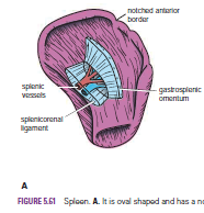
the body. It is oval shaped and is reddish and has a notched anterior border.
It lies just beneath the left half of the diaphragm close to the 9th, 10th, and 11th ribs. The long axis lies along the shaft of the 10th rib, and its lower pole extends forward only as far as the midaxillary line and cannot be palpated on clinical examination .
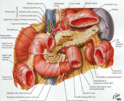
The spleen is surrounded by peritoneum, which passes from it at the hilum as the gastrosplenic omentum (ligament) to the greater curvature of the stomach(carrying the short gastric and left gastroepiploic vessels). The peritoneum also passes to the left kidney as the splenicorenal ligament (carrying the splenic vessels and the tail of the pancreas).
It lies just beneath the left half of the diaphragm close to the 9th, 10th, and 11th ribs. The long axis lies along the shaft of the 10th rib, and its lower pole extends forward only as far as the midaxillary line and cannot be palpated on clinical examination .
The spleen is surrounded by peritoneum, which passes from it at the hilum as the gastrosplenic omentum (ligament) to the greater curvature of the stomach(carrying the short gastric and left gastroepiploic vessels). The peritoneum also passes to the left kidney as the splenicorenal ligament (carrying the splenic vessels and the tail of the pancreas).
Relations
■■
Anteriorly: The stomach, tail of the pancreas, and left colic flexure. The left
kidney lies along its medial border.
■■
Posteriorly: The diaphragm; left pleura (left costodiaphragmatic recess); left
lung; and 9th, 10th, and 11th ribs.

Splenic
Enlargement
An enlarged spleen extends downward and medially. direct
downward enlargement of the organ is prevented by The left colic flexure and
the phrenicocolic ligament . As the enlarged spleen projects below the left
costal margin, its notched anterior border can be recognized by palpation through
the anterior abdominal wall.
The spleen is situated at the beginning of the splenic vein,
and in cases of portal hypertension it often enlarges from venous congestion.
Trauma
to the Spleen
Although anatomically the spleen gives the appearance of being
well protected, automobile accidents of the crushing or run-over type commonly
produce laceration of the spleen. Penetrating wounds of the lower left thorax
can also damage the spleen.