Showing posts with label Hernia. Show all posts

Friday, 17 June 2016

Ascites- Peritoneal Infection-Internal Abdominal Hernia-Peritoneal Dialysis

Ascites
Ascites an excessive accumulation of peritoneal fluid within the peritoneal cavity. it can occur as a result to hepatic cirrhosis (portal venous congestion), malignant disease (e.g., cancer of the testis), or congestive heart failure. In a thin patient, as much as 1500 mL has to accumulate before ascites can be recognized clinically. In obese individuals, a far greater amount has to collect before it can be detected.

Peritoneal Infection
Infection may gain entrance to the peritoneal cavity through several routes: from the interior of the gastrointestinal tract and gallbladder, through the anterior abdominal wall, via the uterine tubes in females (gonococcal peritonitis in adults and pneumococcal peritonitis in children occur through this route), and from the blood. Collection of infected peritoneal fluid in one of the subphrenic spaces is often accompanied by infection of the pleural cavity. It is common to find a localized empyema in a patient with a subphrenic abscess. It is believed that the infection spreads from the peritoneum to the pleura via the diaphragmatic lymph vessels. A patient with a subphrenic abscess may complain of pain over the shoulder. (This also holds true for collections of blood under the diaphragm, which irritate the parietal diaphragmatic peritoneum.) The skin of the shoulder is supplied by the supraclavicular nerves (C3 and 4), which have the same segmental origin as the phrenic nerve, which supplies the peritoneum in the center of the undersurface of the diaphragm. To avoid the accumulation of infected fluid in the subphrenic spaces and to delay the absorption of toxins from intraperitoneal infections, it is common nursing practice to sit a patient up in bed with the back at an angle of 45°. In this position, the infected peritoneal fluid tends to gravitate downward into the pelvic cavity, where the rate of toxin absorption is slow .


Internal Abdominal Hernia
When  a loop of intestine enters a peritoneal pouch or recess like the lesser sac or the duodenal recesses and becomes strangulated at the edges of the recess. Remember that important structures form the boundaries of the entrance into the lesser sac and that the inferior mesenteric vein often lies in the anterior wall of the paraduodenal recess.

Peritoneal Dialysis
Because the peritoneum is a semipermeable membrane, it allows rapid bidirectional transfer of substances across itself. Because the surface area of the peritoneum is enormous, this transfer property has been made use of in patients with acute renal insufficiency. The efficiency of this method is only a fraction of that achieved by hemodialysis.
A watery solution, the dialysate, is introduced through a catheter through a small midline incision through the anterior abdominal wall below the umbilicus. The technique is the same as peritoneal lavage. The products of metabolism, such as urea, diffuse through the peritoneal lining cells from the blood vessels into the dialysate and are removed from the patient.


Umbilical Herniae-Acquired infantile umbilical hernia-Acquired umbilical hernia of adults -Epigastric Hernia-Incisional Hernia-Separation of the Recti Abdominis

Umbilical Herniae
Congenital umbilical hernia, is caused by a failure of part of the midgut to return to the abdominal cavity from the extraembryonic coelom during fetal life.The hernial sac and its relationship to the umbilical cord are shown below:


Acquired infantile umbilical hernia
is a small hernia that sometimes occurs in children and is caused by a weakness in the scar of the umbilicus in the linea alba. Most become smaller and disappear without treatment as the abdominal cavity enlarges.
Acquired umbilical hernia of adults
referred to as a paraumbilical hernia. The hernial sac does not protrude through the umbilical scar, but through the linea alba in the region of the umbilicus. Paraumbilical herniae gradually increase in size and hang downward. The neck of the sac may be narrow, but the body of the sac often contains coils of small and large intestines and omentum. Paraumbilical herniae are much more common in women than in men

Epigastric Hernia
Epigastric hernia occurs through the widest part of the linea alba, anywhere between the xiphoid process and the umbilicus. The hernia is usually small and starts off as a small protrusion of extraperitoneal fat between the fibers of the linea alba. During the following months or years, the fat is forced farther through the linea alba and eventually drags behind it a small peritoneal sac. The body of the sac often contains a small piece of greater omentum. It is common in middle-aged manual workers.

Incisional Hernia
A postoperative incisional hernia is most likely to occur in patients in whom it was necessary to cut one of the segmental nerves supplying the muscles of the anterior abdominal wall; postoperative wound infection with death (necrosis) of the abdominal musculature is also a common cause. The neck of the sac is usually large, and adhesion and strangulation of its contents are rare complications. In very obese individuals, the extent of the abdominal wall weakness is often difficult to assess.


Separation of the Recti Abdominis
Separation of the recti abdominis occurs in elderly multiparous women with weak abdominal muscles .In this condition, the aponeuroses forming the rectus sheath become excessively stretched. When the patient coughs or strains, the recti separate widely, and a large hernial sac, containing abdominal viscera, bulges forward between the medial margins of the recti. This can be corrected by wearing a suitable abdominal belt


Femoral Hernia-

Femoral Hernia

the femoral hernia is more common in women than in men because of a wider pelvis and femoral canal . The neck of the hernial sac lies below and lateral to the pubic tubercle.

The hernial sac passes down the femoral canal, pushing the femoral septum before it. On escaping through the lower end, it expands to form a swelling in the upper part of the thigh deep to the deep fascia. With further expansion, the hernial sac may turn upward to cross the anterior surface of the inguinal ligament.


The neck of the sac always lies below and lateral to the pubic tubercle , which serves to distinguish it from an inguinal hernia. The neck of the sac is narrow and lies at the femoral ring. The ring is related anteriorly to the inguinal ligament, posteriorly to the pectineal ligament and the pubis, medially to the sharp free edge of the lacunar ligament, and laterally to the femoral vein. Because of the presence of these anatomic structures, the neck of the sac is unable to expand. Once an abdominal viscus has passed through the neck into the body of the sac, it may be difficult to push it up and return it to the abdominal cavity (irreducible hernia). Furthermore, after straining or coughing, a piece of bowel may be forced through the neck and its blood vessels may be compressed by the femoral ring, seriously impairing its blood supply (strangulated hernia). A femoral hernia is a dangerous disease and should always be treated surgically.

The hernial sac descends through the femoral canal within the femoral sheath, creating a femoral hernia. The femoral sheath, is a protrusion of the fascial envelope lining the abdominal walls and surrounds the femoral vessels and lymphatics for about 1 in. (2.5 cm) below the inguinal ligament . The femoral artery, as it enters the thigh below the inguinal ligament, occupies the lateral compartment of the sheath. The femoral vein, which lies on its medial side and is separated from it by a fibrous septum, occupies the intermediate compartment. The lymph vessels, which are separated from the vein by a fibrous septum, occupy the most medial compartment. The femoral canal, the compartment for the lymphatics, occupies the medial part of the sheath. It is about 0.5 in. (1.3 cm) long, and its upper opening is referred to as the femoral ring. The femoral septum, which is a condensation of extraperitoneal tissue, plugs the opening of the femoral ring.


Abdominal Herniae-Indirect Inguinal Hernia-Direct Inguinal Hernia

Abdominal Herniae
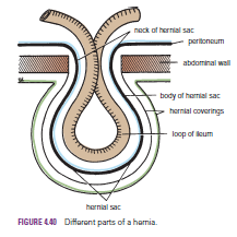
A hernia is the protrusion of part of the abdominal contents beyond the normal confines of the abdominal wall. It consists of three parts: the sac, the contents of the sac, and the coverings of the sac. The hernial sac is a pouch (diverticulum) of peritoneum and has a neck and a body. The hernial contents may consist of any structure found within the abdominal cavity and may vary from a small piece of omentum to a large viscus such as the kidney. The hernial coverings are formed from the layers of the abdominal wall through which the hernial sac passes. Abdominal herniae are of the following common types:

Inguinal (indirect or direct)
 Femoral
Umbilical (congenital or acquired)
 Epigastric
Separation of the recti abdominis
 Incisional
Hernia of the linea semilunaris (Spigelian hernia)
Lumbar

Indirect Inguinal Hernia
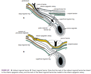
The indirect inguinal hernia is the most common form of hernia It is more common than a direct inguinal hernia. It is much more common in males than females.  and is believed to be congenital in origin . The hernial sac is the remains of the processus vaginalis (an outpouching of peritoneum that in the fetus is responsible for the formation of the inguinal canal . It follows that the sac enters the inguinal canal through the deep inguinal ring lateral to the inferior epigastric vessels. It may extend part of the way along the canal or the full length, as far as the superficial inguinal ring. If the processus vaginalis has undergone no obliteration, then the hernia is complete and extends through the superficial inguinal ring down into the scrotum or labium majus. Under these circumstances, the neck of the hernial sac lies at the deep inguinal ring lateral to the inferior epigastric vessels, and the body of the sac resides in the inguinal canal and scrotum (or base of labium majus). An indirect inguinal hernia is about 20 times more common in males than in females, and nearly one third are bilateral. It is more common on the right (normally, the right processus vaginalis becomes obliterated after the left; the right testis descends later than the left). It is most common in children and young adults


Direct Inguinal Hernia
The direct inguinal hernia It is common in old men with weak abdominal muscles and it makes up about 15% of all inguinal hernias it is rare in women . The sac of a direct hernia bulges directly anteriorly through the posterior wall of the inguinal canal medial to the inferior epigastric vessels  and The neck of the hernial sac is wide. Because of the presence of the strong conjoint tendon (combined tendons of insertion of the internal oblique and transversus muscles), this hernia is usually nothing more than a generalized bulge; therefore, the neck of the hernial sac is wide. Direct inguinal hernias are rare in women and most are bilateral. It is a disease of old men with weak abdominal muscles.