Umbilical
Herniae
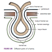
Congenital umbilical hernia, is caused by a failure of part
of the midgut to return to the abdominal cavity from the extraembryonic coelom
during fetal life.The hernial sac and its relationship to the umbilical cord
are shown below:
Acquired infantile umbilical hernia
is a small hernia that sometimes occurs in children and is caused by a weakness in the scar of the umbilicus in the linea alba. Most become smaller and disappear without treatment as the abdominal cavity enlarges.
is a small hernia that sometimes occurs in children and is caused by a weakness in the scar of the umbilicus in the linea alba. Most become smaller and disappear without treatment as the abdominal cavity enlarges.
Acquired umbilical hernia of adults
referred to as a paraumbilical hernia. The hernial sac does not protrude through the umbilical scar, but through the linea alba in the region of the umbilicus. Paraumbilical herniae gradually increase in size and hang downward. The neck of the sac may be narrow, but the body of the sac often contains coils of small and large intestines and omentum. Paraumbilical herniae are much more common in women than in men
referred to as a paraumbilical hernia. The hernial sac does not protrude through the umbilical scar, but through the linea alba in the region of the umbilicus. Paraumbilical herniae gradually increase in size and hang downward. The neck of the sac may be narrow, but the body of the sac often contains coils of small and large intestines and omentum. Paraumbilical herniae are much more common in women than in men
Epigastric
Hernia
Epigastric hernia occurs through the widest part of the
linea alba, anywhere between the xiphoid process and the umbilicus. The hernia
is usually small and starts off as a small protrusion of extraperitoneal fat
between the fibers of the linea alba. During the following months or years, the
fat is forced farther through the linea alba and eventually drags behind it a
small peritoneal sac. The body of the sac often contains a small piece of
greater omentum. It is common in middle-aged manual workers.
Incisional
Hernia
A postoperative incisional hernia is most likely to occur in
patients in whom it was necessary to cut one of the segmental nerves supplying
the muscles of the anterior abdominal wall; postoperative wound infection with
death (necrosis) of the abdominal musculature is also a common cause. The neck of
the sac is usually large, and adhesion and strangulation of its contents are
rare complications. In very obese individuals, the extent of the abdominal wall
weakness is often difficult to assess.
Separation
of the Recti Abdominis
Separation of the recti abdominis occurs in elderly
multiparous women with weak abdominal muscles .In this condition, the
aponeuroses forming the rectus sheath become excessively stretched. When the
patient coughs or strains, the recti separate widely, and a large hernial sac,
containing abdominal viscera, bulges forward between the medial margins of the
recti. This can be corrected by wearing a suitable abdominal belt


