Showing posts with label Inguinal. Show all posts

Friday, 17 June 2016

Abdominal Herniae-Indirect Inguinal Hernia-Direct Inguinal Hernia

Abdominal Herniae
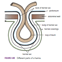
A hernia is the protrusion of part of the abdominal contents beyond the normal confines of the abdominal wall. It consists of three parts: the sac, the contents of the sac, and the coverings of the sac. The hernial sac is a pouch (diverticulum) of peritoneum and has a neck and a body. The hernial contents may consist of any structure found within the abdominal cavity and may vary from a small piece of omentum to a large viscus such as the kidney. The hernial coverings are formed from the layers of the abdominal wall through which the hernial sac passes. Abdominal herniae are of the following common types:

Inguinal (indirect or direct)
 Femoral
Umbilical (congenital or acquired)
 Epigastric
Separation of the recti abdominis
 Incisional
Hernia of the linea semilunaris (Spigelian hernia)
Lumbar

Indirect Inguinal Hernia
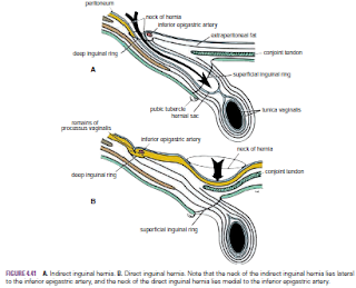
The indirect inguinal hernia is the most common form of hernia It is more common than a direct inguinal hernia. It is much more common in males than females.  and is believed to be congenital in origin . The hernial sac is the remains of the processus vaginalis (an outpouching of peritoneum that in the fetus is responsible for the formation of the inguinal canal . It follows that the sac enters the inguinal canal through the deep inguinal ring lateral to the inferior epigastric vessels. It may extend part of the way along the canal or the full length, as far as the superficial inguinal ring. If the processus vaginalis has undergone no obliteration, then the hernia is complete and extends through the superficial inguinal ring down into the scrotum or labium majus. Under these circumstances, the neck of the hernial sac lies at the deep inguinal ring lateral to the inferior epigastric vessels, and the body of the sac resides in the inguinal canal and scrotum (or base of labium majus). An indirect inguinal hernia is about 20 times more common in males than in females, and nearly one third are bilateral. It is more common on the right (normally, the right processus vaginalis becomes obliterated after the left; the right testis descends later than the left). It is most common in children and young adults


Direct Inguinal Hernia
The direct inguinal hernia It is common in old men with weak abdominal muscles and it makes up about 15% of all inguinal hernias it is rare in women . The sac of a direct hernia bulges directly anteriorly through the posterior wall of the inguinal canal medial to the inferior epigastric vessels  and The neck of the hernial sac is wide. Because of the presence of the strong conjoint tendon (combined tendons of insertion of the internal oblique and transversus muscles), this hernia is usually nothing more than a generalized bulge; therefore, the neck of the hernial sac is wide. Direct inguinal hernias are rare in women and most are bilateral. It is a disease of old men with weak abdominal muscles.


Inguinal Canal- Function of the Inguinal Canal- Walls of the Inguinal Canal-Vas Deferens (Ductus Deferens)

Inguinal Canal
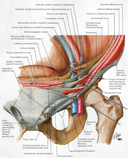
The inguinal canal is an oblique passage through the lower part of the anterior abdominal wall. In the males, it allows structures to pass to and from the testis to the abdomen. In females, it allows the round ligament of the uterus to pass from the uterus to the labium majus. The canal is about 1.5 in. (4 cm) long in the adult and extends from the deep inguinal ring, a hole in the fascia transversalis , downward and medially to the superficial inguinal ring, a hole in the aponeurosis of the external oblique muscle . It lies parallel to and immediately above the inguinal ligament. In the newborn child, the deep ring lies almost directly posterior to the superficial ring so that the canal is considerably shorter at this age. Later, as the result of growth, the deep ring moves laterally. The deep inguinal ring,* an oval opening in the fascia transversalis, lies about 0.5 in. (1.3 cm) above the inguinal ligament midway between the anterior superior iliac spine and the symphysis pubis . Related to it medially are the inferior epigastric vessels, which pass upward from the external iliac vessels. The margins of the ring give attachment to the internal spermatic fascia (or the internal covering of the round ligament of the uterus). The superficial inguinal ring* is a triangular-shaped defect in the aponeurosis of the external oblique muscle and lies immediately above and medial to the pubic tubercle . The margins of the ring, sometimes called the crura, give attachment to the external spermatic fascia.


Function of the Inguinal Canal
The inguinal canal allows structures of the spermatic cord to pass to and from the testis to the abdomen in the male. (Normal spermatogenesis takes place only if the testis leaves the abdominal cavity to enter a cooler environment in the scrotum.) In the female, the smaller canal permits the passage of the round ligament of the uterus from the uterus to the labium majus

Walls of the Inguinal Canal
Anterior wall:
 External oblique aponeurosis, reinforced laterally by the origin of the internal oblique from the inguinal ligament . This wall is therefore strongest where it lies opposite the weakest part of the posterior wall, namely, the deep inguinal ring.
Posterior wall:
 Conjoint tendon medially, fascia transversalis laterally . This wall is therefore strongest where it lies opposite the weakest part of the
anterior wall:
 namely, the superficial inguinal ring. Roof or superior wall. Arching lowest fibers of the internal oblique and transversus abdominis muscles .
Floor or inferior wall:
Upturned lower edge of the inguinal ligament and, at its medial end, the lacunar ligament


Vas Deferens (Ductus Deferens)
The vas deferens is a cordlike structure that can be palpated between finger and thumb in the upper part of the scrotum. It is a thick-walled muscular duct that transports spermatozoa from the epididymis to the urethra.