Showing posts with label Tendon. Show all posts

Wednesday, 29 June 2016

The Wrist and Hand-Important Structures Lying in Front of the Wrist-Radial Artery-Tendon of Flexor Carpi Radialis-Tendon of Palmaris Longus (If Present)-Tendons of Flexor Digitorum Superficialis-Tendon of Flexor Carpi Ulnaris-Ulnar Artery-Ulnar Nerve-Important Structures Lying on the Lateral Side of the Wrist-Anatomic Snuffbox-Important Structures Lying on the Back of the Wrist-Lunate-Important Structures Lying in the Palm-Recurrent Branch of the Median Nerve-Superficial Palmar Arterial Arch-Important Structures Lying on the Dorsum of the Hand

The Wrist and Hand
At the wrist, the styloid processes of the radius and ulna can be palpated. The styloid process of the radius lies about 0.75 in. (1.9 cm) distal to that of the ulna.
The dorsal tubercle of the radius is palpable on the posterior surface of the distal end of the radius.
The head of the ulna is most easily felt with the forearm pronated; the head then stands out prominently on the lateral side of the wrist. The rounded head can be distinguished from the more distal pointed styloid process.
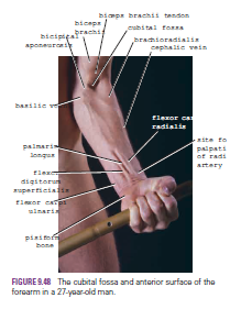
The pisiform bone can be felt on the medial side of the anterior aspect of the wrist between the two transverse creases. The hook of the hamate bone can be felt on deep palpation of the hypothenar eminence, a fingerbreadth distal and lateral to the pisiform bone.
The transverse creases seen in front of the wrist are important landmarks. The proximal transverse crease lies at the level of the wrist joint. The distal transverse crease corresponds to the proximal border of the flexor retinaculum.

Important Structures Lying in Front of the Wrist
Radial Artery
The pulsations of the radial artery can easily be felt anterior to the distal third of the radius. Here, it lies just beneath the skin and fascia lateral to the tendon of flexor carpi radialis muscle
Tendon of Flexor Carpi Radialis
The tendon of the flexor carpi radialis lies medial to the pulsating radial artery
.
Tendon of Palmaris Longus (If Present)
The tendon of the palmaris longus lies medial to the tendon of flexor carpi radialis and overlies the median nerve
Tendons of Flexor Digitorum Superficialis
The tendons of the flexor digitorum superficialis are a group of four that lie medial to the tendon of palmaris longus and can be seen moving beneath the skin when the fingers are flexed and extended.

 
Tendon of Flexor Carpi Ulnaris
The tendon of the flexor carpi ulnaris is the most medially placed tendon on the front of the wrist and can be followed distally to its insertion on the pisiform bone. The tendon can be made prominent by asking the patient to clench the fist (the muscle contracts to assist in fixing and stabilizing the wrist joint)
.
Ulnar Artery
The pulsations of the ulnar artery can be felt lateral to the tendon of flexor carpi ulnaris
Ulnar Nerve
The ulnar nerve lies immediately medial to the ulnar artery


Important Structures Lying on the Lateral Side of the Wrist
Anatomic Snuffbox
The “anatomic snuffbox” is an important area. It is a skin depression that lies distal to the styloid process of the radius. It is bounded medially by the tendon of extensor pollicis longus and laterally by the tendons of abductor pollicis longus and extensor pollicis brevis. In its floor can be palpated the styloid process of the radius (proximally) and the base of the first metacarpal bone of the thumb (distally); between these bones beneath the floor lie the scaphoid and the trapezium (felt but not identifiable).
The radial artery can be palpated within the snuffbox as the artery winds around the lateral margin of the wrist to reach the dorsum of the hand. The cephalic vein can also sometimes be recognized crossing the snuffbox as it ascends the forearm.

Important Structures Lying on the Back of the Wrist
Lunate
The lunate lies in the proximal row of carpal bones. It can be palpated just distal to the dorsal tubercle of the radius when the wrist joint is flexed.


Important Structures Lying in the Palm
Recurrent Branch of the Median Nerve
The recurrent branch to the muscles of the thenar eminence curves around the lower border of the flexor retinaculum and lies about one fingerbreadth distal to the tubercle of the scaphoid
Superficial Palmar Arterial Arch
The superficial palmar arterial arch is located in the central part of the palm and lies on a line drawn across the palm at the level of the distal border of the fully extended thumb
Deep Palmar Arterial Arch
The deep palmar arterial arch is also located in the central part of the palm and lies on a line drawn across the palm at the level of the proximal border of the fully extended thumb
Metacarpophalangeal Joints
The metacarpophalangeal joints lie approximately at the level of the distal transverse palmar crease. The interphalangeal joints lie at the level of the middle and distal finger creases.

Important Structures Lying on the Dorsum of the Hand
The tendons of extensor digitorum, the extensor indicis, and the extensor digiti minimi can be seen and felt as they pass distally to the bases of the fingers.


Sunday, 26 June 2016

Rotator Cuff-Quadrangular Space-Rotator Cuff Tendinitis-Rupture of the Supraspinatus Tendon-Arterial Anastomosis around the Shoulder Joint-Branches from the Subclavian Artery- Branches from the Axillary Artery-Arterial Anastomosis and Ligation of the Axillary Artery

Rotator Cuff
The rotator cuff is the name given to the tendons of the subscapularis, supraspinatus, infraspinatus, and teres minor muscles, which are fused to the underlying capsule of the shoulder joint. The cuff plays a very important role in stabilizing the shoulder joint. The tone of these muscles assists in holding the head of the humerus in the glenoid cavity of the scapula during movements at the shoulder joint. The cuff lies on the anterior, superior, and posterior aspects of the joint. The cuff is deficient inferiorly, and this is a site of potential weakness.

Quadrangular Space
The quadrangular space is an intermuscular space, located immediately below the shoulder joint. It is bounded above by the subscapularis and capsule of the shoulder joint and below by the teres major muscle. It is bounded medially by the long head of the triceps and laterally by the surgical neck of the humerus.
The axillary nerve and the posterior circumflex humeral vessels pass backward through this space .

Rotator Cuff Tendinitis
The rotator cuff, consisting of the tendons of the subscapularis, supraspinatus, infraspinatus, and teres minor muscles, which are fused to the underlying capsule of the shoulder joint, plays an important role in stabilizing the shoulder joint. The rotator cuff presses the humeral head into the glenoid cavity. Lesions of the cuff are a common cause of pain in the shoulder region.
Failure of the cuff is due to either wear or tear. Wear is age related. Excessive overhead activity of the upper limb may be the cause of tendinitis, although many cases appear spontaneously. During abduction of the shoulder joint, the supraspinatus tendon is exposed to friction against the acromion. Under normal conditions, the amount of friction is reduced to a minimum by the large subacromial bursa, which extends laterally beneath the deltoid. Degenerative changes in the bursa are followed by degenerative changes in the underlying supraspinatus tendon, and these may extend into the other tendons of the rotator cuff. Clinically, the condition is known as subacromial bursitis, supraspinatus tendinitis, or pericapsulitis. It is characterized by the presence of a spasm of pain in the middle range of abduction, when the diseased area impinges on the acromion. Extensive acute traumatic tears are best repaired surgically as soon as possible. Small chronic cuff injuries are best managed without surgery using nonsteroidal anti-inflammatory drugs and muscle exercises.

Rupture of the Supraspinatus Tendon
In advanced cases of rotator cuff tendinitis, the necrotic supraspinatus tendon can become calcified or rupture. Rupture of the tendon seriously interferes with the normal abduction movement of the shoulder joint. It will be remembered that the main function of the supraspinatus muscle is to hold the head of the humerus in the glenoid fossa at the commencement of abduction. The patient with a ruptured supraspinatus tendon is unable to initiate abduction of the arm. However, if the arm is passively assisted for the first 15° of abduction, the deltoid can then take over and complete the movement to a right angle
.
Arterial Anastomosis around the Shoulder Joint
The extreme mobility of the shoulder joint may result in kinking of the axillary artery and a temporary occlusion of its lumen. To compensate for this, an important arterial anastomosis exists between the branches of the subclavian artery and the axillary artery, thus ensuring that an adequate blood flow takes place into the upper limb irrespective of the position of the arm
Branches from the Subclavian Artery
■■ The suprascapular artery, which is distributed to the supraspinous and infraspinous fossae of the scapula
■■ The superficial cervical artery, which gives off a deep branch that runs down the medial border of the scapula

 
Branches from the Axillary Artery
■■ The subscapular artery and its circumflex scapular branch supply the subscapular and infraspinous fossae of the scapula, respectively.
■■ The anterior circumflex humeral artery
■■ The posterior circumflex humeral artery Both the circumflex arteries form an anastomosing circle around the surgical neck of the humerus.

Arterial Anastomosis and Ligation of the Axillary Artery
The existence of the anastomosis around the shoulder joint is vital to preserving the upper limb should it be necessary to ligate the axillary artery.