The
Wrist and Hand
At the wrist, the styloid processes of the radius and ulna
can be palpated. The styloid process of the radius lies about 0.75 in. (1.9 cm)
distal to that of the ulna.
The dorsal tubercle of the radius is palpable on the
posterior surface of the distal end of the radius.
The head of the ulna is most easily felt with the forearm pronated;
the head then stands out prominently on the lateral side of the wrist. The
rounded head can be distinguished from the more distal pointed styloid process.
The pisiform bone can be felt on the medial side of the
anterior aspect of the wrist between the two transverse creases. The hook of
the hamate bone can be felt on deep palpation of the hypothenar eminence, a
fingerbreadth distal and lateral to the pisiform bone.
The transverse creases seen in front of the wrist are important
landmarks. The proximal transverse crease lies at the level of the wrist joint.
The distal transverse crease corresponds to the proximal border of the flexor
retinaculum.
Radial Artery
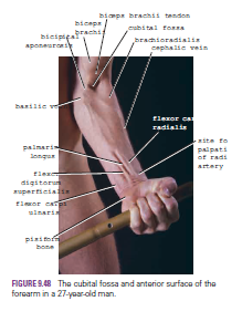
The pulsations of the radial artery can easily be felt
anterior to the distal third of the radius. Here, it lies just beneath the skin
and fascia lateral to the tendon of flexor carpi radialis muscle
Tendon of Flexor Carpi Radialis
The tendon of the flexor carpi radialis lies medial to the pulsating radial artery
.
The tendon of the flexor carpi radialis lies medial to the pulsating radial artery
.
Tendon of Palmaris Longus (If Present)
The tendon of the palmaris longus lies medial to the tendon of
flexor carpi radialis and overlies the median nerve
Tendons of Flexor Digitorum Superficialis
The tendons of the flexor digitorum superficialis are a group
of four that lie medial to the tendon of palmaris longus and can be seen moving
beneath the skin when the fingers are flexed and extended.
Tendon of Flexor Carpi Ulnaris
The tendon of the flexor carpi ulnaris is the most medially placed
tendon on the front of the wrist and can be followed distally to its insertion
on the pisiform bone. The tendon can be made prominent by asking the patient to
clench the fist (the muscle contracts to assist in fixing and stabilizing the
wrist joint)
.
.
Ulnar Artery
The pulsations of the ulnar artery can be felt lateral to
the tendon of flexor carpi ulnaris
Ulnar Nerve
The ulnar nerve lies immediately medial to the ulnar artery
Important Structures Lying on the Lateral Side of the Wrist
Anatomic Snuffbox
The “anatomic snuffbox” is an important area. It is a skin depression
that lies distal to the styloid process of the radius. It is bounded medially
by the tendon of extensor pollicis longus and laterally by the tendons of
abductor pollicis longus and extensor pollicis brevis. In its floor can be
palpated the styloid process of the radius (proximally) and the base of the
first metacarpal bone of the thumb (distally); between these bones beneath the
floor lie the scaphoid and the trapezium (felt but not identifiable).
The radial artery can be palpated within the snuffbox as the
artery winds around the lateral margin of the wrist to reach the dorsum of the
hand. The cephalic vein can also sometimes be recognized crossing the snuffbox as
it ascends the forearm.
Important
Structures Lying on the Back of the Wrist
Lunate
The lunate lies in the proximal row of carpal bones. It can be
palpated just distal to the dorsal tubercle of the radius when the wrist joint
is flexed.
Important
Structures Lying in the Palm
Recurrent Branch of the Median Nerve
The recurrent branch to the muscles of the thenar eminence curves
around the lower border of the flexor retinaculum and lies about one
fingerbreadth distal to the tubercle of the scaphoid
Superficial Palmar Arterial Arch
The superficial palmar arterial arch is located in the
central part of the palm and lies on a line drawn across the palm at the level
of the distal border of the fully extended thumb
Deep Palmar Arterial Arch
The deep palmar arterial arch is also located in the central
part of the palm and lies on a line drawn across the palm at the level of the
proximal border of the fully extended thumb
Metacarpophalangeal Joints
The metacarpophalangeal joints lie approximately at the level
of the distal transverse palmar crease. The interphalangeal joints lie at the
level of the middle and distal finger creases.
Important
Structures Lying on the Dorsum of the Hand
The tendons of extensor digitorum, the extensor indicis, and
the extensor digiti minimi can be seen and felt as they pass distally to the
bases of the fingers.


