Showing posts with label Artery. Show all posts

Sunday, 28 August 2016

Arteries of the Gluteal Region-Superior Gluteal Artery-Inferior Gluteal Artery-The- Trochanteric Anastomosis

Arteries of the Gluteal Region
Superior Gluteal Artery
The superior gluteal artery is a branch from the internal iliac artery and enters the gluteal region through the upper part of the greater sciatic foramen above the piriformis. It divides into branches that are distributed throughout the gluteal region.

 
Inferior Gluteal Artery
The inferior gluteal artery is a branch of the internal iliac artery and enters the gluteal region through the lower part of the greater sciatic foramen, below the piriformis. It divides into numerous branches that are distributed throughout the gluteal region.

The Trochanteric Anastomosis
The trochanteric anastomosis provides the main blood supply to the head of the femur. The nutrient arteries pass along the femoral neck beneath the capsule. The following arteries take part in the anastomosis: the superior gluteal artery, the inferior gluteal artery, the medial femoral circumflex artery, and the lateral femoral circumflex artery.

The Cruciate Anastomosis
The cruciate anastomosis is situated at the level of the lesser trochanter of the femur and, together with the trochanteric anastomosis, provides a connection between the internal iliac and the femoral arteries. The following arteries take part in the anastomosis: the inferior gluteal artery, the medial femoral circumflex artery, the lateral femoral circumflex artery, and the first perforating artery, a branch of the profunda artery.

Veins of the Lower Limb
The veins of the lower limb can be divided into three groups: superficial, deep, and perforating. The superficial veins consist of the great and small saphenous veins and their tributaries, which are situated beneath the skin in the superficial fascia.
The constant position of the great saphenous vein in front of the medial malleolus should be remembered for patients requiring emergency blood transfusion. The deep veins are the venae comitantes to the anterior and posterior tibial arteries, the popliteal vein, and the femoral veins and their tributaries. The perforating veins are communicating vessels that run between the superficial and deep veins. Many of these veins are found particularly in the region of the ankle and the medial side of the lower part of the leg. They possess valves that are arranged to prevent the flow of blood from the deep to the superficial veins.


Wednesday, 29 June 2016

The Wrist and Hand-Important Structures Lying in Front of the Wrist-Radial Artery-Tendon of Flexor Carpi Radialis-Tendon of Palmaris Longus (If Present)-Tendons of Flexor Digitorum Superficialis-Tendon of Flexor Carpi Ulnaris-Ulnar Artery-Ulnar Nerve-Important Structures Lying on the Lateral Side of the Wrist-Anatomic Snuffbox-Important Structures Lying on the Back of the Wrist-Lunate-Important Structures Lying in the Palm-Recurrent Branch of the Median Nerve-Superficial Palmar Arterial Arch-Important Structures Lying on the Dorsum of the Hand

The Wrist and Hand
At the wrist, the styloid processes of the radius and ulna can be palpated. The styloid process of the radius lies about 0.75 in. (1.9 cm) distal to that of the ulna.
The dorsal tubercle of the radius is palpable on the posterior surface of the distal end of the radius.
The head of the ulna is most easily felt with the forearm pronated; the head then stands out prominently on the lateral side of the wrist. The rounded head can be distinguished from the more distal pointed styloid process.
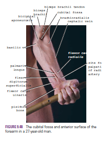
The pisiform bone can be felt on the medial side of the anterior aspect of the wrist between the two transverse creases. The hook of the hamate bone can be felt on deep palpation of the hypothenar eminence, a fingerbreadth distal and lateral to the pisiform bone.
The transverse creases seen in front of the wrist are important landmarks. The proximal transverse crease lies at the level of the wrist joint. The distal transverse crease corresponds to the proximal border of the flexor retinaculum.

Important Structures Lying in Front of the Wrist
Radial Artery
The pulsations of the radial artery can easily be felt anterior to the distal third of the radius. Here, it lies just beneath the skin and fascia lateral to the tendon of flexor carpi radialis muscle
Tendon of Flexor Carpi Radialis
The tendon of the flexor carpi radialis lies medial to the pulsating radial artery
.
Tendon of Palmaris Longus (If Present)
The tendon of the palmaris longus lies medial to the tendon of flexor carpi radialis and overlies the median nerve
Tendons of Flexor Digitorum Superficialis
The tendons of the flexor digitorum superficialis are a group of four that lie medial to the tendon of palmaris longus and can be seen moving beneath the skin when the fingers are flexed and extended.

 
Tendon of Flexor Carpi Ulnaris
The tendon of the flexor carpi ulnaris is the most medially placed tendon on the front of the wrist and can be followed distally to its insertion on the pisiform bone. The tendon can be made prominent by asking the patient to clench the fist (the muscle contracts to assist in fixing and stabilizing the wrist joint)
.
Ulnar Artery
The pulsations of the ulnar artery can be felt lateral to the tendon of flexor carpi ulnaris
Ulnar Nerve
The ulnar nerve lies immediately medial to the ulnar artery


Important Structures Lying on the Lateral Side of the Wrist
Anatomic Snuffbox
The “anatomic snuffbox” is an important area. It is a skin depression that lies distal to the styloid process of the radius. It is bounded medially by the tendon of extensor pollicis longus and laterally by the tendons of abductor pollicis longus and extensor pollicis brevis. In its floor can be palpated the styloid process of the radius (proximally) and the base of the first metacarpal bone of the thumb (distally); between these bones beneath the floor lie the scaphoid and the trapezium (felt but not identifiable).
The radial artery can be palpated within the snuffbox as the artery winds around the lateral margin of the wrist to reach the dorsum of the hand. The cephalic vein can also sometimes be recognized crossing the snuffbox as it ascends the forearm.

Important Structures Lying on the Back of the Wrist
Lunate
The lunate lies in the proximal row of carpal bones. It can be palpated just distal to the dorsal tubercle of the radius when the wrist joint is flexed.


Important Structures Lying in the Palm
Recurrent Branch of the Median Nerve
The recurrent branch to the muscles of the thenar eminence curves around the lower border of the flexor retinaculum and lies about one fingerbreadth distal to the tubercle of the scaphoid
Superficial Palmar Arterial Arch
The superficial palmar arterial arch is located in the central part of the palm and lies on a line drawn across the palm at the level of the distal border of the fully extended thumb
Deep Palmar Arterial Arch
The deep palmar arterial arch is also located in the central part of the palm and lies on a line drawn across the palm at the level of the proximal border of the fully extended thumb
Metacarpophalangeal Joints
The metacarpophalangeal joints lie approximately at the level of the distal transverse palmar crease. The interphalangeal joints lie at the level of the middle and distal finger creases.

Important Structures Lying on the Dorsum of the Hand
The tendons of extensor digitorum, the extensor indicis, and the extensor digiti minimi can be seen and felt as they pass distally to the bases of the fingers.


Tuesday, 14 June 2016

Coronary Artery Disease-Arteriosclerotic disease of the coronary arteries


Coronary Artery Disease
The myocardium receives its blood supply through the right and left coronary arteries. Although the coronary arteries have numerous anastomoses at the arteriolar level, they are essentially functional end arteries. A sudden block of one of the large branches of either coronary artery will usually lead to necrosis of the cardiac muscle (myocardial infarction) in that vascular area, and often the patient dies. Most cases of coronary artery blockage are caused by an acute thrombosis on top of a chronic atherosclerotic narrowing of the lumen


Arteriosclerotic disease of the coronary arteries

it may present in three ways, depending on the rate of narrowing of the lumina of the arteries: (1) General degeneration and fibrosis of the myocardium occur over many years and are caused by a gradual narrowing of the coronary arteries. (2) Angina pectoris is cardiac pain that occurs on exertion and is relieved by rest. In this condition, the coronary arteries are so narrowed that myocardial ischemia occurs on exertion but not at rest. (3) Myocardial infarction occurs when coronary flow is suddenly reduced or stopped and the cardiac muscle undergoes necrosis. Myocardial infarction is the major cause of death in industrialized nations.

Because coronary bypass surgery, coronary angioplasty, and coronary artery stenting are now commonly accepted methods of treating coronary artery disease, it is incumbent on the student to be prepared to interpret still- and motion-picture angiograms that have been carried out before treatment. For this reason, a working knowledge of the origin, course, and distribution of the coronary arteries should be memorized.


Monday, 13 June 2016

Internal Thoracic Artery in the Treatment of Coronary Artery Disease- Lymph Drainage of the Thoracic Wall

Internal Thoracic Artery in the Treatment of Coronary Artery Disease
In patients with occlusive coronary disease caused by atherosclerosis, the diseased arterial segment can be bypassed by inserting a graft. The graft most commonly used is the great saphenous vein of the leg. In some patients, the myocardium can be revascularized by surgically mobilizing one of the internal thoracic arteries and joining its distal cut end to a coronary artery.


 Lymph Drainage of the Thoracic Wall

 The lymph drainage of the skin of the anterior chest wall passes to the anterior axillary lymph nodes; that from the posterior chest wall passes to the posterior axillary nodes. The lymph drainage of the intercostal spaces passes forward to the internal thoracic nodes, situated along the internal thoracic artery, and posteriorly to the posterior intercostal nodes and the para-aortic nodes in the posterior mediastinum.