Axillary
Nerve
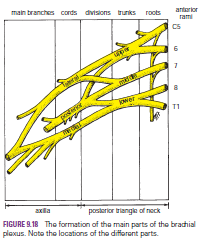
The axillary nerve, which arises from the posterior cord of
the brachial plexus (C5 and 6), can be injured by the pressure of a badly
adjusted crutch pressing upward into the armpit.
The passage of the axillary nerve backward from the axilla through
the quadrangular space makes it particularly vulnerable here to downward
displacement of the humeral head in shoulder dislocations or fractures of the
surgical neck of the humerus.
Paralysis of the deltoid and teres minor muscles results. The
cutaneous branches of the axillary nerve, including the upper lateral cutaneous
nerve of the arm, are functionless, and consequently there is a loss of skin
sensation over the lower half of the deltoid muscle. The paralyzed deltoid
wastes rapidly, and the underlying greater tuberosity can be readily palpated.
Because the supraspinatus is the only other abductor of the shoulder, this movement
is much impaired. Paralysis of the teres minor is not recognizable clinically.
Radial
Nerve
The radial nerve, which arises from the posterior cord of
the brachial plexus, characteristically gives off its branches some distance
proximal to the part to be innervated.
In the axilla, it gives off three branches: the posterior
cutaneous nerve of the arm, which supplies the skin on the back of the arm down
to the elbow; the nerve to the long head of the triceps; and the nerve to the
medial head of the triceps.
In the spiral groove of the humerus, it gives off four
branches: the lower lateral cutaneous nerve of the arm, which supplies the lateral
surface of the arm down to the elbow; the posterior cutaneous nerve of the
forearm, which supplies the skin down the middle of the back of the forearm as
far as the wrist; the nerve to the lateral head of the triceps; and the nerve
to the medial head of the triceps and the anconeus.
In the anterior compartment of the arm above the lateral
epicondyle, it gives off three branches: the nerve to a small part of the brachialis,
the nerve to the brachioradialis, and the nerve to the extensor carpi radialis
longus.
In the cubital fossa, it gives off the deep branch of the
radial nerve and continues as the superficial radial nerve. The deep branch
supplies the extensor carpi radialis brevis and the supinator in the cubital
fossa and all the extensor muscles in the posterior compartment of the forearm.
The superficial radial nerve is sensory and supplies the skin over the lateral
part of the dorsum of the hand and the dorsal surface of the lateral three and
a half fingers proximal to the nail beds. (The ulnar nerve supplies the medial
part of the dorsum of the hand and the dorsal surface of the medial one and a
half fingers; the exact cutaneous areas innervated by the radial and ulnar
nerves on the hand are subject to variation.)
The radial nerve is commonly damaged in the axilla and in
the spiral groove
Musculocutaneous
Nerve
The musculocutaneous nerve is rarely injured because of its
protected position beneath the biceps brachii muscle. If it is injured high up
in the arm, the biceps and coracobrachialis are paralyzed and the brachialis
muscle is weakened (the latter muscle is also supplied by the radial nerve).
Flexion of the forearm at the elbow joint is then produced by the remainder of
the brachialis muscle and the flexors of the forearm. When the forearm is in
the prone position, the extensor carpi radialis longus and the brachioradialis
muscles assist in flexion of the forearm.
There is also sensory loss along the lateral side of the
forearm. Wounds or cuts of the forearm can sever the lateral cutaneous nerve of
the forearm, a continuation of the musculocutaneous nerve beyond the cubital
fossa, resulting in sensory loss along the lateral side of the forearm.
Median
Nerve
The median nerve, which arises from the medial and lateral
cords of the brachial plexus, gives off no cutaneous or motor branches in the
axilla or in the arm. In the proximal third of the front of the forearm, by
unnamed branches or by its anterior interosseous branch, it supplies all the
muscles of the front of the forearm except the flexor carpi ulnaris and the
medial half of the flexor digitorum profundus, which are supplied by the ulnar nerve.
In the distal third of the forearm, it gives rise to a palmar cutaneous branch,
which crosses in front of the flexor retinaculum and supplies the skin on the
lateral half of the palm In the palm, the median nerve supplies the muscles of
the thenar eminence and the first two lumbricals and gives sensory innervation
to the skin of the palmar aspect of the lateral three and a half fingers,
including the nail beds on the dorsum.
From a clinical standpoint, the median nerve is injured
occasionally in the elbow region in supracondylar fractures of the humerus. It
is most commonly injured by stab wounds or broken glass just proximal to the
flexor retinaculum; here, it lies in the interval between the tendons of the
flexor carpi radialis and flexor digitorum superficialis, overlapped by the
palmaris longus.


