Showing posts with label a. Show all posts

Sunday, 26 June 2016

Brachial Plexus-The Axillary Sheath and a Brachial Plexus Nerve Block

Brachial Plexus
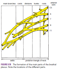
The nerves entering the upper limb provide the following important functions: sensory innervation to the skin and deep structures, such as the joints; motor innervation to the muscles; influence over the diameters of the blood vessels by the sympathetic vasomotor nerves; and sympathetic secretomotor supply to the sweat glands. At the root of the neck, the nerves form a complicated plexus called the brachial plexus. This allows the nerve fibers derived from different segments of the spinal cord to be arranged and distributed efficiently in different nerve trunks to the various parts of the upper limb. The brachial plexus is formed in the posterior triangle of the neck by the union of the anterior rami of the 5th, 6th, 7th, and 8th cervical and the 1st thoracic spinal nerves.
The plexus can be divided into roots, trunks, divisions, and cords. The roots of C5 and 6 unite to form the upper trunk, the root of C7 continues as the middle trunk, and the roots of C8 and T1 unite to form the lower trunk. Each trunk then divides into anterior and posterior divisions. The anterior divisions of the upper and middle trunks unite to form the lateral cord, the anterior division of the lower trunk continues as the medial cord, and the posterior divisions of all three trunks join to form the posterior cord.
The roots, trunks, and divisions of the brachial plexus reside in the lower part of the posterior triangle of the neck and are fully described on page XXX. The cords become arranged around the axillary artery in the axilla. Here, the brachial plexus and the axillary artery and vein are enclosed in the axillary sheath.
Cords of the Brachial Plexus All three cords of the brachial plexus lie above and lateral to the first part of the axillary artery. The medial cord crosses behind the artery to reach the medial side of the second part of the artery . The posterior cord lies behind the second part of the artery, and the lateral cord lies on the lateral side of the second part of the artery . Thus, the cords of the plexus have the relationship to the second part of the axillary artery that is indicated by their names.


Most branches of the cords that form the main nerve trunks of the upper limb continue this relationship to the artery in its third part .
The branches of the different parts of the brachial plexus are as follows:
■■ Roots
Dorsal scapular nerve (C5)
Long thoracic nerve (C5, 6, and 7)

■■ Upper trunk
Nerve to subclavius (C5 and 6)
Suprascapular nerve (supplies the supraspinatus and
infraspinatus muscles)
■■ Lateral cord
Lateral pectoral nerve
Musculocutaneous nerve
Lateral root of median nerve
■■ Medial cord
Medial pectoral nerve
Medial cutaneous nerve of arm and medial cutaneous
nerve of forearm
Ulnar nerve
Medial root of median nerve
■■ Posterior cord
Upper and lower subscapular nerves
Thoracodorsal nerve
Axillary nerve
Radial nerve
The Axillary Sheath and a Brachial Plexus Nerve Block
Because the axillary sheath encloses the axillary vessels and the brachial plexus, a brachial plexus nerve block can easily be obtained. The distal part of the sheath is closed with finger pressure, and a syringe needle is inserted into the proximal part of the sheath. The anesthetic solution is then injected into the sheath, and the solution is massaged along the sheath to produce the nerve block. The position of the sheath can be verified by feeling the pulsations of the third part of the axillary artery.


Tuesday, 21 June 2016

Uterine Tube-Uterine TubeFunction-Blood Supply-The Uterine Tube as a Conduit for Infection-Pelvic Inflammatory Disease-Ectopic Pregnancy- Tubal Ligation-

Uterine Tube
The two uterine tubes are each about 4 in. (10 cm) long and lie in the upper border of the broad ligament . Each connects the peritoneal cavity in the region of the ovary with the cavity of the uterus. The uterine tube is divided into four parts:
1. The infundibulum is the funnel-shaped lateral end that projects beyond the broad ligament and overlies the ovary. The free edge of the funnel has several fingerlike processes, known as fimbriae, which are draped over the ovary .
2. The ampulla is the widest part of the tube.
3. The isthmus is the narrowest part of the tube and lies just lateral to the uterus .
4. The intramural part is the segment that pierces the uterine wall .

Function
The uterine tube receives the ovum from the ovary and provides a site where fertilization of the ovum can take place (usually in the ampulla). It provides nourishment for the fertilized ovum and transports it to the cavity of the uterus. The tube serves as a conduit along which the spermatozoa travel to reach the ovum.

Blood Supply

Arteries
The uterine artery from the internal iliac artery and the ovarian artery from the abdominal aorta .

Veins
The veins correspond to the arteries.

Lymph Drainage
The internal iliac and para-aortic nodes.


Nerve Supply
Sympathetic and parasympathetic nerves from the inferior hypogastric plexuses

The Uterine Tube as a Conduit for Infection
The uterine tube lies in the upper free border of the broad ligament and is a direct route of communication from the vulva through the vagina and uterine cavity to the peritoneal cavity.

Pelvic Inflammatory Disease
The pathogenic organism(s) enter the body through sexual contact and ascend through the uterus and enter the uterine tubes. Salpingitis may follow, with leakage of pus into the peritoneal cavity, causing pelvic peritonitis. A pelvic abscess usually follows, or the infection spreads farther, causing general peritonitis.

Ectopic Pregnancy
Implantation and growth of a fertilized ovum may occur outside the uterine cavity in the wall of the uterine tube. This is a variety of ectopic pregnancy. There being no decidua formation in the tube, the eroding action of the trophoblast quickly destroys the wall of the tube. Tubal abortion or rupture of the tube, with the effusion of a large quantity of blood into the peritoneal cavity, is the common result.
The blood pours down into the rectouterine pouch (pouch of Douglas) or into the uterovesical pouch. The blood may quickly ascend into the general peritoneal cavity, giving rise to severe abdominal pain, tenderness, and guarding. Irritation of the subdiaphragmatic peritoneum (supplied by phrenic nerves C3, 4, and 5) may give rise to referred pain to the shoulder skin (supraclavicular nerves C3 and 4).


Tubal Ligation
Ligation and division of the uterine tubes is a method of obtaining permanent birth control and is usually restricted to women who already have children. The ova that are discharged from the ovarian follicles degenerate in the tube proximal to the obstruction. If, later, the woman wishes to have an additional child, restoration of the continuity of the uterine tubes can be attempted, and, in about 20% of women, fertilization occurs.