Showing posts with label Plexus. Show all posts

Sunday, 3 July 2016

Brachial Plexus Injuries-Lower Lesions of the Brachial Plexus (Klumpke Palsy)-Long Thoracic Nerve

Brachial Plexus Injuries
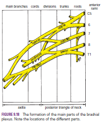
The roots, trunks, and divisions of the brachial plexus reside in the lower part of the posterior triangle of the neck, whereas the cords and most of the branches of the plexus lie in the axilla.
Complete lesions involving all the roots of the plexus are rare.
Incomplete injuries are common and are usually caused by traction or pressure; individual nerves can be divided by stab wounds.
Upper Lesions of the Brachial Plexus (Erb–Duchenne Palsy) Upper lesions of the brachial plexus are injuries resulting from excessive displacement of the head to the opposite side and depression of the shoulder on the same side. This causes excessive traction or even tearing of C5 and 6 roots of the plexus. It occurs in infants during a difficult delivery or in adults after a blow to or fall on the shoulder. The suprascapular nerve, the nerve to the subclavius, and the musculocutaneous and axillary nerves all possess nerve fibers derived from C5 and 6 roots and will therefore be functionless. The following muscles will consequently be paralyzed: the supraspinatus (abductor of the shoulder) and infraspinatus (lateral rotator of the shoulder); the subclavius (depresses the clavicle); the biceps brachii (supinator of the forearm, flexor of the elbow, weak flexor of the shoulder) and the greater part of the brachialis (flexor of the elbow) and the coracobrachialis (flexes the shoulder); and the deltoid (abductor of the shoulder) and the teres minor (lateral rotator of the shoulder).
Thus, the limb will hang limply by the side, medially rotated by the unopposed sternocostal part of the pectoralis major; the forearm will be pronated because of loss of the action of the biceps. The position of the upper limb in this condition has been likened to that of a porter or waiter hinting for a tip. In addition, there will be a loss of sensation down the lateral side of the arm.

Lower Lesions of the Brachial Plexus (Klumpke Palsy)
Lower lesions of the brachial plexus are usually traction injuries caused by excessive abduction of the arm, as occurs in the case of a person falling from a height clutching at an object to save himself or herself. The 1st thoracic nerve is usually torn.
The nerve fibers from this segment run in the ulnar and median nerves to supply all the small muscles of the hand. The hand has a clawed appearance caused by hyperextension of the metacarpophalangeal joints and flexion of the interphalangeal joints. The extensor digitorum is unopposed by the lumbricals and interossei and extends the metacarpophalangeal joints; the flexor digitorum superficialis and profundus are unopposed by the lumbricals and interossei and flex the middle and terminal phalanges, respectively.

In addition, loss of sensation will occur along the medial side of the arm. If the 8th cervical nerve is also damaged, the extent of anesthesia will be greater and will involve the medial side of the forearm, hand, and medial two fingers.
Lower lesions of the brachial plexus can also be produced by the presence of a cervical rib or malignant metastases from the lungs in the lower deep cervical lymph nodes.

Long Thoracic Nerve
The long thoracic nerve, which arises from C5, 6, and 7 and supplies the serratus anterior muscle, can be injured by blows to or pressure on the posterior triangle of the neck or during the surgical procedure of radical mastectomy. Paralysis of the serratus anterior results in the inability to rotate the scapula during the movement of abduction of the arm above a right angle.
The patient therefore experiences difficulty in raising the arm above the head. The vertebral border and inferior angle of the scapula will no longer be kept closely applied to the chest wall and will protrude posteriorly, a condition known as “winged scapula”


Sunday, 26 June 2016

Brachial Plexus-The Axillary Sheath and a Brachial Plexus Nerve Block

Brachial Plexus
The nerves entering the upper limb provide the following important functions: sensory innervation to the skin and deep structures, such as the joints; motor innervation to the muscles; influence over the diameters of the blood vessels by the sympathetic vasomotor nerves; and sympathetic secretomotor supply to the sweat glands. At the root of the neck, the nerves form a complicated plexus called the brachial plexus. This allows the nerve fibers derived from different segments of the spinal cord to be arranged and distributed efficiently in different nerve trunks to the various parts of the upper limb. The brachial plexus is formed in the posterior triangle of the neck by the union of the anterior rami of the 5th, 6th, 7th, and 8th cervical and the 1st thoracic spinal nerves.
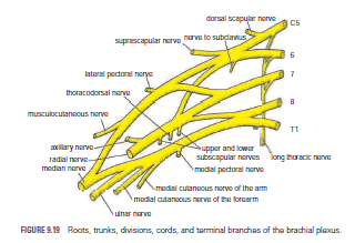
The plexus can be divided into roots, trunks, divisions, and cords. The roots of C5 and 6 unite to form the upper trunk, the root of C7 continues as the middle trunk, and the roots of C8 and T1 unite to form the lower trunk. Each trunk then divides into anterior and posterior divisions. The anterior divisions of the upper and middle trunks unite to form the lateral cord, the anterior division of the lower trunk continues as the medial cord, and the posterior divisions of all three trunks join to form the posterior cord.
The roots, trunks, and divisions of the brachial plexus reside in the lower part of the posterior triangle of the neck and are fully described on page XXX. The cords become arranged around the axillary artery in the axilla. Here, the brachial plexus and the axillary artery and vein are enclosed in the axillary sheath.
Cords of the Brachial Plexus All three cords of the brachial plexus lie above and lateral to the first part of the axillary artery. The medial cord crosses behind the artery to reach the medial side of the second part of the artery . The posterior cord lies behind the second part of the artery, and the lateral cord lies on the lateral side of the second part of the artery . Thus, the cords of the plexus have the relationship to the second part of the axillary artery that is indicated by their names.


Most branches of the cords that form the main nerve trunks of the upper limb continue this relationship to the artery in its third part .
The branches of the different parts of the brachial plexus are as follows:
■■ Roots
Dorsal scapular nerve (C5)
Long thoracic nerve (C5, 6, and 7)

■■ Upper trunk
Nerve to subclavius (C5 and 6)
Suprascapular nerve (supplies the supraspinatus and
infraspinatus muscles)
■■ Lateral cord
Lateral pectoral nerve
Musculocutaneous nerve
Lateral root of median nerve
■■ Medial cord
Medial pectoral nerve
Medial cutaneous nerve of arm and medial cutaneous
nerve of forearm
Ulnar nerve
Medial root of median nerve
■■ Posterior cord
Upper and lower subscapular nerves
Thoracodorsal nerve
Axillary nerve
Radial nerve
The Axillary Sheath and a Brachial Plexus Nerve Block
Because the axillary sheath encloses the axillary vessels and the brachial plexus, a brachial plexus nerve block can easily be obtained. The distal part of the sheath is closed with finger pressure, and a syringe needle is inserted into the proximal part of the sheath. The anesthetic solution is then injected into the sheath, and the solution is massaged along the sheath to produce the nerve block. The position of the sheath can be verified by feeling the pulsations of the third part of the axillary artery.


Sunday, 19 June 2016

Sacral Plexus-Pressure from the Fetal Head-Invasion by Malignant Tumors-Referred Pain from the Obturator Nerve-Caudal Anesthesia (Analgesia)

Sacral Plexus
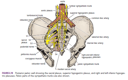
The sacral plexus lies in front of the piriformis muscle on the posterior pelvic wall. It is formed from the anterior rami of the 4th and 5th lumbar nerves and the anterior rami of the first, second, third, and fourth sacral nerves. The fourth lumbar nerve joins the fifth lumbar nerve to form the lumbosacral trunk. The lumbosacral trunk passes down into the pelvis and joins the sacral nerves as they emerge from the anterior sacral foramina.

 
Pressure from the Fetal Head
when the fetal head has descended into the pelvis During the later stages of pregnancy, , the mother often complains of discomfort or aching pain extending down one of the lower limbs. The discomfort, caused by pressure from the fetal head, is often relieved by changing position, such as lying on the side in bed.

Invasion by Malignant Tumors
The nerves of the sacral plexus can become invaded by malignant tumors extending from neighboring viscera. A carcinoma of the rectum, for example, can cause severe intractable pain down the lower limbs.

Referred Pain from the Obturator Nerve
The obturator nerve lies on the lateral wall of the pelvis and supplies the parietal peritoneum. An inflamed appendix hanging down into the pelvic cavity could cause irritation of the obturator nerve endings, leading to referred pain down the inner side of the right thigh. Inflammation of the ovaries can produce similar symptoms.

Caudal Anesthesia (Analgesia)
Anesthetic solutions can be injected into the sacral canal through the sacral hiatus. The solutions then act on the spinal roots of the 2nd, 3rd, 4th and 5th sacral and coccygeal segments of the cord as they emerge from the dura mater. The roots of higher spinal segments can also be blocked by this method. The needle must be confined to the lower part of the sacral canal, because the meninges extend down as far as the lower border of the second sacral vertebra. Caudal anesthesia is used in obstetrics to block pain fibers from the cervix of the uterus and to anesthetize the perineum