Showing posts with label Function. Show all posts

Wednesday, 29 June 2016

Movements of the Thumb-Diseases of the Hand and Preservation of Function-

Movements of the Thumb
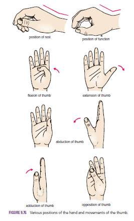
Flexion is the movement of the thumb across the palm in such a manner as to maintain the plane of the thumbnail at right angles to the plane of the other fingernails.
The movement takes place between the trapezium and the1st metacarpal bone, at the metacarpophalangeal and interphalangeal joints. The muscles producing the movement are the flexor pollicis longus and brevis and the opponens pollicis.
Extension is the movement of the thumb in a lateral or coronal plane away from the palm in such a manner as to maintain the plane of the thumbnail at right angles to the plane of the other fingernails. The movement takes place between the trapezium and the 1st metacarpal bone, at the metacarpophalangeal and interphalangeal joints. The muscles producing the movement are the extensor pollicis longus and brevis.
Abduction is the movement of the thumb in an anteroposterior plane away from the palm, the plane of the thumbnail being kept at right angles to the plane of the other nails. The movement takes place mainly between the trapezium and the 1st metacarpal bone; a small amount of movement takes place at the metacarpophalangeal joint. The muscles producing the movement are the abductor pollicis longus and brevis.
Adduction is the movement of the thumb in an anteroposterior plane toward the palm, the plane of the thumbnail being kept at right angles to the plane of the other fingernails. The movement takes place between the trapezium and the 1st metacarpal bone.
The muscle producing the movement is the adductor pollicis.

Opposition is the movement of the thumb across the palm in such a manner that the anterior surface of the tip comes into contact with the anterior surface of the tip of any of the other fingers. The movement is accomplished by the medial rotation of the 1st metacarpal bone and the attached phalanges on the trapezium.
The plane of the thumbnail comes to lie parallel with the plane of the nail of the opposed finger. The muscle producing the movement is the opponens pollicis.
 
Diseases of the Hand and Preservation of Function
From the clinical standpoint, the hand is one of the most important organs of the body. Without a normally functioning hand, the patient’s livelihood is often in jeopardy. To students who doubt this statement, I would suggest that they place their right (or left) hand in a pocket for 24 hours. They will be astonished at the number of times they would like to use it if they could.
From the purely mechanical point of view, the hand can be regarded as a pincer-like mechanism between the thumb and fingers, situated at the end of a multijointed lever. The most important part of the hand is the thumb, and it is the physician’s responsibility to preserve the thumb, or as much of it as possible, so that the pincer-like mechanism can be maintained. The pincer- like action of the thumb largely depends on its unique ability to be drawn across the palm and opposed to the other fingers.
This movement alone, although important, is insufficient for the mechanism to work effectively. The opposing skin surfaces must have tactile sensation—and this explains why median nerve palsy is so much more disabling than ulnar nerve palsy.
If the hand requires immobilization for the treatment of disease of any part of the upper limb, it should be immobilized (if possible) in the position of function. This means that if loss of movement occurs at the wrist joint, or at the joints of the hand or fingers, the patient will at least have a hand that is in a position of mechanical advantage, and one that can serve a useful purpose.
Physicians should also remember that when a finger (excluding the thumb) is normally flexed into the palm, it points to the tubercle of the scaphoid; individual fingers requiring immobilization in flexion, on a splint or within a cast, should therefore always be placed in this position.
Always refer to the patient’s fingers by name: thumb, index, middle, ring, and little finger. Numbering the fingers is confusing (is the thumb a finger?) and has led to such disastrous results as amputating the wrong finger.


Friday, 24 June 2016

Vagina-Relations-Function-Supports of the Vagina-Vaginal Examination-Prolapse of the Vagina-Vaginal Trauma

Vagina
The vagina is a muscular tube that extends upward and backward from the vulva to the uterus. It measures about 3 in. (8 cm) long and has anterior and posterior walls, which are normally in apposition. At its upper end, the anterior wall is pierced by the cervix, which projects downward and backward into the vagina. It is important to remember that the upper half of the vagina lies above the pelvic floor and the lower half lies within the perineum. The area of the vaginal lumen, which surrounds the cervix, is divided into four regions, or fornices: anterior, posterior, right lateral, and left lateral. The vaginal orifice in a virgin possesses a thin mucosal fold called the hymen, which is perforated at its center. After childbirth, the hymen usually consists only of tags

Relations
■■ Anteriorly: The vagina is closely related to the bladder above and to the urethra below.
■■ Posteriorly: The upper third of the vagina is related to the rectouterine pouch (pouch of Douglas) and its middle third to the ampulla of the rectum. The lower third is related to the perineal body, which separates it from the anal canal.

■■ Laterally: In its upper part, the vagina is related to the ureter; its middle part is related to the anterior fibers of the levator ani, as they run backward to reach the perineal body and hook around the anorectal junction. Contraction of the fibers of levator ani compresses the walls of the vagina together. In its lower part, the vagina is related to the urogenital diaphragm and the bulb of the vestibule.

 
Function
The vagina not only is the female genital canal, but it also serves as the excretory duct for the menstrual flow and

Supports of the Vagina
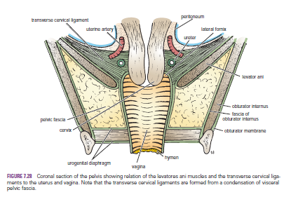
The upper part of the vagina is supported by the levatores ani muscles and the transverse cervical, pubocervical, and sacrocervical ligaments. These structures are attached to the vaginal wall by pelvic fascia. The middle part of the vagina is supported by the urogenital diaphragm. The lower part of the vagina, especially the posterior wall, is supported by the perineal body.

Vaginal Examination
The anatomic relations of the vagina are of great clinical importance. Many pathologic conditions occurring in the female pelvis may be diagnosed using a simple vaginal examination.
The following structures can be palpated through the vaginal walls from above downward:
■■ Anteriorly: The bladder and the urethra
■■ Posteriorly: Loops of ileum and the sigmoid colon in the rectouterine peritoneal pouch (pouch of Douglas), the rectal ampulla, and the perineal body
■■ Laterally: The ureters, the pelvic fascia and the anterior fibers of the levatores ani muscles, and the urogenital diaphragm

Prolapse of the Vagina
The vaginal vault is supported by the same structures that support the uterine cervix. Prolapse of the uterus is necessarily associated with some degree of sagging of the vaginal walls. However, if the supports of the bladder, urethra, or anterior rectal wall are damaged in childbirth, prolapse of the vaginal walls occurs, with the uterus remaining in its correct position.
Sagging of the bladder results in the bulging of the anterior wall of the vagina, a condition known as a cystocele. When the ampulla of the rectum sags against the posterior vaginal wall, the bulge is called a rectocele.

Vaginal Trauma
Coital injury, picket fence–type of impalement injury, and vaginal perforation caused by water under pressure, as occurs in water skiing, are common injuries. Lacerations of the vaginal wall involving the posterior fornix may violate the pouch of Douglas of the peritoneal cavity and cause prolapse of the small intestine into the vagina

forms part of the birth canal.


Uterus-Uterus Relations-Function- Positions of the Uterus-Supports of the Uterus-The Levatores Ani Muscles and the Perineal Body-

Uterus
The uterus is a hollow, pear-shaped organ with thick muscular walls. In the young nulliparous adult, it measures 3 in. (8 cm) long, 2 in. (5 cm) wide, and 1 in. (2.5 cm) thick. It is divided into the fundus, body, and cervix.
The fundus is the part of the uterus that lies above the entrance of the uterine tubes.
The body is the part of the uterus that lies below the entrance of the uterine tubes.
The cervix is the narrow part of the uterus. It pierces the anterior wall of the vagina and is divided into the supravaginal and vaginal parts of the cervix. The cavity of the uterine body is triangular in coronal section, but it is merely a cleft in the sagittal plane. The cavity of the cervix, the cervical canal, communicates with the cavity of the body through the internal os and with that of the vagina through the external os. Before the birth of the first child, the external os is circular. In a parous woman, the vaginal part of the cervix is larger, and the external os becomes a transverse slit so that it possesses an anterior lip and a posterior lip.

 
Relations
■■ Anteriorly: The body of the uterus is related anteriorly to the uterovesical pouch and the superior surface of the bladder. The supravaginal cervix is related to the superior surface of the bladder. The vaginal cervix is related to the anterior fornix of the vagina.
■■ Posteriorly: The body of the uterus is related posteriorly to the rectouterine pouch (pouch of Douglas) with coils of ileum or sigmoid colon within it.

■■ Laterally: The body of the uterus is related laterally to the broad ligament and the uterine artery and vein. The supravaginal cervix is related to the ureter as it passes forward to enter the bladder. The vaginal cervix is related to the lateral fornix of the vagina. The uterine tubes enter the superolateral angles of the uterus, and the round ligaments of the ovary and of the uterus are attached to the uterine wall just below this level.

 
Function
The uterus serves as a site for the reception, retention, and nutrition of the fertilized ovum.

Positions of the Uterus
In most women, the long axis of the uterus is bent forward on the long axis of the vagina. This position is referred to as anteversion of the uterus. Furthermore, the long axis of the body of the uterus is bent forward at the level of the internal os with the long axis of the cervix. This position is termed anteflexion of the uterus. Thus, in the erect position and with the bladder empty, the uterus lies in an almost horizontal plane. In some women, the fundus and body of the uterus are bent backward on the vagina so that they lie in the rectouterine pouch (pouch of Douglas). In this situation, the uterus is said to be retroverted. If the body of the uterus is, in addition, bent backward on the cervix, it is said to be retroflexed

Supports of the Uterus
The uterus is supported mainly by the tone of the levatores ani muscles and the condensations of pelvic fascia, which form three important ligaments.

The Levatores Ani Muscles and the Perineal Body
 They form a broad muscular sheet stretching across the pelvic cavity, and, together with the pelvic fascia on their upper surface, they effectively support the pelvic viscera and resist the intra-abdominal pressure transmitted downward through the pelvis. The medial edges of the anterior parts of the levatores ani muscles are attached to the cervix of the uterus by the pelvic fascia.


Tuesday, 21 June 2016

Uterine Tube-Uterine TubeFunction-Blood Supply-The Uterine Tube as a Conduit for Infection-Pelvic Inflammatory Disease-Ectopic Pregnancy- Tubal Ligation-

Uterine Tube
The two uterine tubes are each about 4 in. (10 cm) long and lie in the upper border of the broad ligament . Each connects the peritoneal cavity in the region of the ovary with the cavity of the uterus. The uterine tube is divided into four parts:
1. The infundibulum is the funnel-shaped lateral end that projects beyond the broad ligament and overlies the ovary. The free edge of the funnel has several fingerlike processes, known as fimbriae, which are draped over the ovary .
2. The ampulla is the widest part of the tube.
3. The isthmus is the narrowest part of the tube and lies just lateral to the uterus .
4. The intramural part is the segment that pierces the uterine wall .

Function
The uterine tube receives the ovum from the ovary and provides a site where fertilization of the ovum can take place (usually in the ampulla). It provides nourishment for the fertilized ovum and transports it to the cavity of the uterus. The tube serves as a conduit along which the spermatozoa travel to reach the ovum.

Blood Supply

Arteries
The uterine artery from the internal iliac artery and the ovarian artery from the abdominal aorta .

Veins
The veins correspond to the arteries.

Lymph Drainage
The internal iliac and para-aortic nodes.


Nerve Supply
Sympathetic and parasympathetic nerves from the inferior hypogastric plexuses

The Uterine Tube as a Conduit for Infection
The uterine tube lies in the upper free border of the broad ligament and is a direct route of communication from the vulva through the vagina and uterine cavity to the peritoneal cavity.

Pelvic Inflammatory Disease
The pathogenic organism(s) enter the body through sexual contact and ascend through the uterus and enter the uterine tubes. Salpingitis may follow, with leakage of pus into the peritoneal cavity, causing pelvic peritonitis. A pelvic abscess usually follows, or the infection spreads farther, causing general peritonitis.

Ectopic Pregnancy
Implantation and growth of a fertilized ovum may occur outside the uterine cavity in the wall of the uterine tube. This is a variety of ectopic pregnancy. There being no decidua formation in the tube, the eroding action of the trophoblast quickly destroys the wall of the tube. Tubal abortion or rupture of the tube, with the effusion of a large quantity of blood into the peritoneal cavity, is the common result.
The blood pours down into the rectouterine pouch (pouch of Douglas) or into the uterovesical pouch. The blood may quickly ascend into the general peritoneal cavity, giving rise to severe abdominal pain, tenderness, and guarding. Irritation of the subdiaphragmatic peritoneum (supplied by phrenic nerves C3, 4, and 5) may give rise to referred pain to the shoulder skin (supraclavicular nerves C3 and 4).


Tubal Ligation
Ligation and division of the uterine tubes is a method of obtaining permanent birth control and is usually restricted to women who already have children. The ova that are discharged from the ovarian follicles degenerate in the tube proximal to the obstruction. If, later, the woman wishes to have an additional child, restoration of the continuity of the uterine tubes can be attempted, and, in about 20% of women, fertilization occurs.