Showing posts with label Lower. Show all posts

Sunday, 28 August 2016

Venous Pump of the Lower Limb-Varicose Veins

Venous Pump of the Lower Limb
Within the closed fascial compartments of the lower limb, the thinwalled, valved venae comitantes are subjected to intermittent pressure at rest and during exercise. The pulsations of the adjacent arteries help move the blood up the limb. However, the contractions of the large muscles within the compartments during exercise compress these deeply placed veins and force the blood up the limb.
The superficial saphenous veins, except near their termination, lie within the superficial fascia and are not subject to these compression forces. The valves in the perforating veins prevent the high-pressure venous blood from being forced outward into the low-pressure superficial veins. Moreover, as the muscles within the closed fascial compartments relax, venous blood is sucked from the superficial into the deep veins.

Varicose Veins
A varicosed vein is one that has a larger diameter than normal and is elongated and tortuous. Varicosity of the esophageal and rectal veins is described elsewhere.
This condition commonly occurs in the superficial veins of the lower limb and, although not life threatening, is responsible for considerable discomfort and pain.
Varicosed veins have many causes, including hereditary weakness of the vein walls and incompetent valves; elevated intraabdominal pressure as a result of multiple pregnancies or abdominal tumors; and thrombophlebitis of the deep veins, which results in the superficial veins becoming the main venous pathway for the lower limb. It is easy to understand how this condition can be produced by incompetence of a valve in a perforating vein. Every time the patient exercises, high-pressure venous blood escapes from the deep veins into the superficial veins and produces a varicosity, which might be localized to begin with but becomes more extensive later. The successful operative treatment of varicosed veins depends on the ligation and division of all the main tributaries of the great or small saphenous veins, to prevent a collateral venous circulation from developing, and the ligation and division of all the perforating veins responsible for the leakage of highpressure blood from the deep to the superficial veins. It is now common practice to remove or strip the superficial veins in addition.
Needless to say, it is imperative to ascertain that the deep veins are patent before operative measures are taken.


Sunday, 3 July 2016

Brachial Plexus Injuries-Lower Lesions of the Brachial Plexus (Klumpke Palsy)-Long Thoracic Nerve

Brachial Plexus Injuries
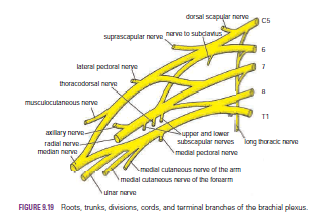
The roots, trunks, and divisions of the brachial plexus reside in the lower part of the posterior triangle of the neck, whereas the cords and most of the branches of the plexus lie in the axilla.
Complete lesions involving all the roots of the plexus are rare.
Incomplete injuries are common and are usually caused by traction or pressure; individual nerves can be divided by stab wounds.
Upper Lesions of the Brachial Plexus (Erb–Duchenne Palsy) Upper lesions of the brachial plexus are injuries resulting from excessive displacement of the head to the opposite side and depression of the shoulder on the same side. This causes excessive traction or even tearing of C5 and 6 roots of the plexus. It occurs in infants during a difficult delivery or in adults after a blow to or fall on the shoulder. The suprascapular nerve, the nerve to the subclavius, and the musculocutaneous and axillary nerves all possess nerve fibers derived from C5 and 6 roots and will therefore be functionless. The following muscles will consequently be paralyzed: the supraspinatus (abductor of the shoulder) and infraspinatus (lateral rotator of the shoulder); the subclavius (depresses the clavicle); the biceps brachii (supinator of the forearm, flexor of the elbow, weak flexor of the shoulder) and the greater part of the brachialis (flexor of the elbow) and the coracobrachialis (flexes the shoulder); and the deltoid (abductor of the shoulder) and the teres minor (lateral rotator of the shoulder).
Thus, the limb will hang limply by the side, medially rotated by the unopposed sternocostal part of the pectoralis major; the forearm will be pronated because of loss of the action of the biceps. The position of the upper limb in this condition has been likened to that of a porter or waiter hinting for a tip. In addition, there will be a loss of sensation down the lateral side of the arm.

Lower Lesions of the Brachial Plexus (Klumpke Palsy)
Lower lesions of the brachial plexus are usually traction injuries caused by excessive abduction of the arm, as occurs in the case of a person falling from a height clutching at an object to save himself or herself. The 1st thoracic nerve is usually torn.
The nerve fibers from this segment run in the ulnar and median nerves to supply all the small muscles of the hand. The hand has a clawed appearance caused by hyperextension of the metacarpophalangeal joints and flexion of the interphalangeal joints. The extensor digitorum is unopposed by the lumbricals and interossei and extends the metacarpophalangeal joints; the flexor digitorum superficialis and profundus are unopposed by the lumbricals and interossei and flex the middle and terminal phalanges, respectively.

In addition, loss of sensation will occur along the medial side of the arm. If the 8th cervical nerve is also damaged, the extent of anesthesia will be greater and will involve the medial side of the forearm, hand, and medial two fingers.
Lower lesions of the brachial plexus can also be produced by the presence of a cervical rib or malignant metastases from the lungs in the lower deep cervical lymph nodes.

Long Thoracic Nerve
The long thoracic nerve, which arises from C5, 6, and 7 and supplies the serratus anterior muscle, can be injured by blows to or pressure on the posterior triangle of the neck or during the surgical procedure of radical mastectomy. Paralysis of the serratus anterior results in the inability to rotate the scapula during the movement of abduction of the arm above a right angle.
The patient therefore experiences difficulty in raising the arm above the head. The vertebral border and inferior angle of the scapula will no longer be kept closely applied to the chest wall and will protrude posteriorly, a condition known as “winged scapula”


Wednesday, 15 June 2016

Esophageal Constrictions-Carcinoma of the Lower Third of the Esophagus

Esophageal Constrictions
The esophagus has three anatomic and physiologic constrictions. The first is where the pharynx joins the upper end, the second is where the aortic arch and the left bronchus cross its anterior surface, and the third occurs where the esophagus passes through the diaphragm into the stomach. These constrictions are of considerable clinical importance because they are sites where swallowed foreign bodies can lodge or through which it may be difficult to pass an esophagoscope. Because a slight delay in the passage of food or fluid occurs at these levels, strictures develop here after the drinking of caustic fluids. Those constrictions are also the common sites of carcinoma of the esophagus. It is useful to remember that their respective distances from the upper incisor teeth are 6 in. (15 cm), 10 in. (25 cm), and 16 in. (41 cm), respectively


Carcinoma of the Lower Third of the Esophagus
The lymph drainage of the lower third of the esophagus descends through the esophageal opening in the diaphragm and ends in the celiac nodes around the celiac artery A malignant tumor of this area of the esophagus would therefore tend to spread below the diaphragm along this route. Consequently, surgical removal of the lesion would include not only the primary lesion, but also the celiac lymph nodes and all regions that drain into these nodes, namely, the stomach, the upper half of the duodenum, the spleen, and the omenta. Restoration of continuity of the gut is accomplished by performing an esophagojejunostomy