Showing posts with label Injuries. Show all posts

Sunday, 3 July 2016

Injuries to the Median Nerve at the Wrist-Carpal Tunnel Syndrome-Injuries to the Median Nerve at the Elbow-Motor-Sensory-Vasomotor Changes-Trophic Changes-

Injuries to the Median Nerve at the Wrist
■■ Motor: The muscles of the thenar eminence are paralyzed and wasted so that the eminence becomes flattened. The thumb is laterally rotated and adducted. The hand looks flattened and “apelike.” Opposition movement of the thumb is impossible. The first two lumbricals are paralyzed, which can be recognized clinically when the patient is asked to make a fist slowly, and the index and middle fingers tend to lag behind the ring and little fingers.
■■ Sensory, vasomotor, and trophic changes: These changes are identical to those found in the elbow lesions.
Perhaps the most serious disability of all in median nerve injuries is the loss of the ability to oppose the thumb to the other fingers and the loss of sensation over the lateral fingers. The delicate pincer-like action of the hand is no longer possible
Carpal Tunnel Syndrome
The carpal tunnel, formed by the concave anterior surface of the carpal bones and closed by the flexor retinaculum, is tightly packed with the long flexor tendons of the fingers, with their surrounding synovial sheaths, and the median nerve. Clinically, the syndrome consists of a burning pain or “pins and needles” along the distribution of the median nerve to the lateral three and a half fingers and weakness of the thenar muscles. It is produced by compression of the median nerve within the tunnel. The exact cause of the compression is difficult to determine, but thickening of the synovial sheaths of the flexor tendons or arthritic changes in the carpal bones are thought to be responsible in many cases.
As you would expect, no paresthesia occurs over the thenar eminence because this area of skin is supplied by the palmar cutaneous branch of the median nerve, which passes superficially to the flexor retinaculum. The condition is dramatically relieved by decompressing the tunnel by making a longitudinal incision through the flexor retinaculum.

Injuries to the Median Nerve at the Elbow
Motor
The pronator muscles of the forearm and the long flexor muscles of the wrist and fingers, with the exception of the flexor carpi ulnaris and the medial half of the flexor digitorum profundus, will be paralyzed. As a result, the forearm is kept in the supine position; wrist flexion is weak and is accompanied by adduction.
The latter deviation is caused by the paralysis of the flexor carpi radialis and the strength of the flexor carpi ulnaris and the medial half of the flexor digitorum profundus. No flexion is possible at the interphalangeal joints of the index and middle fingers, although weak flexion of the metacarpophalangeal joints of these fingers is attempted by the interossei. When the patient tries to make a fist, the index and to a lesser extent the middle fingers tend to remain straight, whereas the ring and little fingers flex. The latter two fingers are, however, weakened by the loss of the flexor digitorum superficialis.
Flexion of the terminal phalanx of the thumb is lost because of paralysis of the flexor pollicis longus. The muscles of the thenar eminence are paralyzed and wasted so that the eminence is flattened. The thumb is laterally rotated and adducted. The hand looks flattened and “apelike.”

Sensory
Skin sensation is lost on the lateral half or less of the palm of the hand and the palmar aspect of the lateral three and a half fingers.
Sensory loss also occurs on the skin of the distal part of the dorsal surfaces of the lateral three and a half fingers. The area of total anesthesia is considerably less because of the overlap of adjacent nerves.

Vasomotor Changes
The skin areas involved in sensory loss are warmer and drier than normal because of the arteriolar dilatation and absence of sweating resulting from loss of sympathetic control.

Trophic Changes
In long-standing cases, changes are found in the hand and fingers. The skin is dry and scaly, the nails crack easily, and atrophy of the pulp of the fingers is present.

Brachial Plexus Injuries-Lower Lesions of the Brachial Plexus (Klumpke Palsy)-Long Thoracic Nerve

Brachial Plexus Injuries
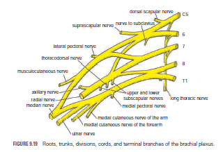
The roots, trunks, and divisions of the brachial plexus reside in the lower part of the posterior triangle of the neck, whereas the cords and most of the branches of the plexus lie in the axilla.
Complete lesions involving all the roots of the plexus are rare.
Incomplete injuries are common and are usually caused by traction or pressure; individual nerves can be divided by stab wounds.
Upper Lesions of the Brachial Plexus (Erb–Duchenne Palsy) Upper lesions of the brachial plexus are injuries resulting from excessive displacement of the head to the opposite side and depression of the shoulder on the same side. This causes excessive traction or even tearing of C5 and 6 roots of the plexus. It occurs in infants during a difficult delivery or in adults after a blow to or fall on the shoulder. The suprascapular nerve, the nerve to the subclavius, and the musculocutaneous and axillary nerves all possess nerve fibers derived from C5 and 6 roots and will therefore be functionless. The following muscles will consequently be paralyzed: the supraspinatus (abductor of the shoulder) and infraspinatus (lateral rotator of the shoulder); the subclavius (depresses the clavicle); the biceps brachii (supinator of the forearm, flexor of the elbow, weak flexor of the shoulder) and the greater part of the brachialis (flexor of the elbow) and the coracobrachialis (flexes the shoulder); and the deltoid (abductor of the shoulder) and the teres minor (lateral rotator of the shoulder).
Thus, the limb will hang limply by the side, medially rotated by the unopposed sternocostal part of the pectoralis major; the forearm will be pronated because of loss of the action of the biceps. The position of the upper limb in this condition has been likened to that of a porter or waiter hinting for a tip. In addition, there will be a loss of sensation down the lateral side of the arm.

Lower Lesions of the Brachial Plexus (Klumpke Palsy)
Lower lesions of the brachial plexus are usually traction injuries caused by excessive abduction of the arm, as occurs in the case of a person falling from a height clutching at an object to save himself or herself. The 1st thoracic nerve is usually torn.
The nerve fibers from this segment run in the ulnar and median nerves to supply all the small muscles of the hand. The hand has a clawed appearance caused by hyperextension of the metacarpophalangeal joints and flexion of the interphalangeal joints. The extensor digitorum is unopposed by the lumbricals and interossei and extends the metacarpophalangeal joints; the flexor digitorum superficialis and profundus are unopposed by the lumbricals and interossei and flex the middle and terminal phalanges, respectively.

In addition, loss of sensation will occur along the medial side of the arm. If the 8th cervical nerve is also damaged, the extent of anesthesia will be greater and will involve the medial side of the forearm, hand, and medial two fingers.
Lower lesions of the brachial plexus can also be produced by the presence of a cervical rib or malignant metastases from the lungs in the lower deep cervical lymph nodes.

Long Thoracic Nerve
The long thoracic nerve, which arises from C5, 6, and 7 and supplies the serratus anterior muscle, can be injured by blows to or pressure on the posterior triangle of the neck or during the surgical procedure of radical mastectomy. Paralysis of the serratus anterior results in the inability to rotate the scapula during the movement of abduction of the arm above a right angle.
The patient therefore experiences difficulty in raising the arm above the head. The vertebral border and inferior angle of the scapula will no longer be kept closely applied to the chest wall and will protrude posteriorly, a condition known as “winged scapula”


Wednesday, 29 June 2016

Wrist Joint (Radiocarpal Joint)-Movements-Important Relations-Wrist Joint Injuries-Falls on the Outstretched Hand-

Wrist Joint (Radiocarpal Joint)
■■ Articulation: Between the distal end of the radius and the articular disc above and the scaphoid, lunate, and triquetral bones below. The proximal articular surface forms an ellipsoid concave surface, which is adapted to the distal ellipsoid convex surface.
■■ Type: Synovial ellipsoid joint
■■ Capsule: The capsule encloses the joint and is attached above to the distal ends of the radius and ulna and below to the proximal row of carpal bones.
■■ Ligaments: Anterior and posterior ligaments strengthen the capsule. The medial ligament is attached to the styloid process of the ulna and to the triquetral bone. The lateral ligament is attached to the styloid process of the radius and to the scaphoid bone.
■■ Synovial membrane: This lines the capsule and is attached to the margins of the articular surfaces. The joint cavity does not communicate with that of the distal radioulnar joint or with the joint cavities of the intercarpal joints.
■■ Nerve supply: Anterior interosseous nerve and the deep branch of the radial nerve

Movements
The following movements are possible: flexion, extension, abduction, adduction, and circumduction. Rotation is not possible because the articular surfaces are ellipsoid shaped.
The lack of rotation is compensated for by the movements of pronation and supination of the forearm.
Flexion is performed by the flexor carpi radialis, the flexor carpi ulnaris, and the palmaris longus. These muscles are assisted by the flexor digitorum superficialis, the flexor digitorum profundus, and the flexor pollicis longus.
Extension is performed by the extensor carpi radialis longus, the extensor carpi radialis brevis, and the extensor carpi ulnaris. These muscles are assisted by the extensor digitorum, the extensor indicis, the extensor digiti minimi, and the extensor pollicis longus.
Abduction is performed by the flexor carpi radialis and the extensor carpi radialis longus and brevis. These muscles are assisted by the abductor pollicis longus and extensor pollicis longus and brevis.
Adduction is performed by the flexor and extensor carpi ulnaris.

Important Relations
■■ Anteriorly: The tendons of the flexor digitorum profundus and superficialis, the flexor pollicis longus, the flexor carpi radialis, the flexor carpi ulnaris, and the median and ulnar nerves
■■ Posteriorly: The tendons of the extensor carpi ulnaris, the extensor digiti minimi, the extensor digitorum, the extensor indicis, the extensor carpi radialis longus and brevis, the extensor pollicis longus and brevis, and the abductor pollicis longus
■■ Medially: The posterior cutaneous branch of the ulnar nerve
■■ Laterally: The radial artery

Wrist Joint Injuries
The wrist joint is essentially a synovial joint between the distal end of the radius and the proximal row of carpal bones. The head of the ulna is separated from the carpal bones by the strong triangular fibrocartilaginous ligament, which separates the wrist joint from the distal radioulnar joint. The joint is stabilized by the strong medial and lateral ligaments.
Because the styloid process of the radius is longer than that of the ulna, abduction of the wrist joint is less extensive than adduction. In flexion–extension movements, the hand can be flexed about 80° but extended to only about 45°. The range of flexion is increased by movement at the midcarpal joint.

A fall on the outstretched hand can strain the anterior ligament of the wrist joint, producing synovial effusion, joint pain, and limitation of movement. These symptoms and signs must not be confused with those produced by a fractured scaphoid or dislocation of the lunate bone, which are similar.

Falls on the Outstretched Hand
In falls on the outstretched hand, forces are transmitted from the scaphoid to the distal end of the radius, from the radius across the interosseous membrane to the ulna, and from the ulna to the humerus; thence, through the glenoid fossa of the scapula to the coracoclavicular ligament and the clavicle; and finally, to the sternum. If the forces are excessive, different parts of the upper limb give way under the strain. The area affected seems to be related to age. In a young child, for example, there may be a posterior displacement of the distal radial epiphysis; in the teenager the clavicle might fracture; in the young adult the scaphoid is commonly fractured; and in the elderly the distal end of the radius is fractured about 1 in. (2.5 cm) proximal to the wrist joint (Colles’ fracture).


Tuesday, 28 June 2016

Injuries to the Bones of the Hand-Dislocation of the lunate bone-Fractures of the metacarpal bones-Bennett’s fracture-Compartment Syndrome of the Forearm--Volkmann’s Ischemic Contracture

Injuries to the Bones of the Hand
Fracture of the scaphoid bone is common in young adults; unless treated effectively, the fragments will not unite, and permanent weakness and pain of the wrist will result, with the subsequent development of osteoarthritis. The fracture line usually goes through the narrowest part of the bone, which, because of its location, is bathed in synovial fluid. The blood vessels to the scaphoid enter its proximal and distal ends, although the blood supply is occasionally confined to its distal end. If the latter occurs, a fracture deprives the proximal fragment of its arterial supply, and this fragment undergoes avascular necrosis.
Deep tenderness in the anatomic snuffbox after a fall on the outstretched hand in a young adult makes one suspicious of a fractured scaphoid.

Dislocation of the lunate bone
occasionally occurs in young adults who fall on the outstretched hand in a way that causes hyperextension of the wrist joint. Involvement of the median nerve is common.

Fractures of the metacarpal bones
 can occur as a result of direct violence, such as the clenched fist striking a hard object.
The fracture always angulates dorsally. The “boxer’s fracture” commonly produces an oblique fracture of the neck of the fifth and sometimes the fourth metacarpal bones. The distal fragment is commonly displaced proximally, thus shortening the finger posteriorly.

Bennett’s fracture
is a fracture of the base of the metacarpal of the thumb caused when violence is applied along the long axis of the thumb or the thumb is forcefully abducted. The fracture is oblique and enters the carpometacarpal joint of the thumb, causing joint instability.
Fractures of the phalanges are common and usually follow direct injury.


Compartment Syndrome of the Forearm
The forearm is enclosed in a sheath of deep fascia, which is attached to the periosteum of the posterior subcutaneous border of the ulna. This fascial sheath, together with the interosseous membrane and fibrous intermuscular septa, divides the forearm into several compartments, each having its own muscles, nerves, and blood supply. There is very little room within each compartment, and any edema can cause secondary vascular compression of the blood vessels; the veins are first affected, and later the arteries.
Soft tissue injury is a common cause, and early diagnosis is critical. Early signs include altered skin sensation (caused by ischemia of the sensory nerves passing through the compartment),
pain disproportionate to any injury (caused by pressure on nerves within the compartment), pain on passive stretching of muscles that pass through the compartment (caused by muscle ischemia), tenderness of the skin over the compartment (a late sign caused by edema), and absence of capillary refill in the nail beds (caused by pressure on the arteries within the compartment). Once the diagnosis is made, the deep fascia must be incised surgically to decompress the affected compartment. A delay of as little as4 hours can cause irreversible damage to the muscles.


Volkmann’s Ischemic Contracture
Volkmann’s ischemic contracture is a contracture of the muscles of the forearm that commonly follows fractures of the distal end of the humerus or fractures of the radius and ulna. In this syndrome, a localized segment of the brachial artery goes into spasm, reducing the arterial flow to the flexor and the extensor muscles so that they undergo ischemic necrosis. The flexor muscles are larger than the extensor muscles, and they are therefore the ones mainly affected. The muscles are replaced by fibrous tissue, which contracts, producing the deformity. The arterial spasm is usually caused by an overtight cast, but in some cases the fracture itself may be responsible. The deformity can be explained only by understanding the anatomy of the region.

Three types of deformity exist:
■■ The long flexor muscles of the carpus and fingers are more contracted than the extensor muscles, and the wrist joint is flexed; the fingers are extended. If the wrist joint is extended passively, the fingers become flexed.
■■ The long extensor muscles to the fingers, which are inserted into the extensor expansion that is attached to the proximal phalanx, are greatly contracted; the metacarpophalangeal joints and the wrist joint are extended, and the interphalangeal joints of the fingers are flexed.
■■ Both the flexor and extensor muscles of the forearm are contracted. The wrist joint is flexed, the metacarpophalangeal joints are extended, and the interphalangeal joints are flexed.