Showing posts with label Pelvis. Show all posts

Sunday, 19 June 2016

Sex Differences of the Pelvis-Pelvic Joints Changes-Changes with Pregnancy-Changes with Age-Sacroiliac Joint Disease-

Sex Differences of the Pelvis
The sex differences of the bony pelvis are easily recognized.
The more obvious differences result from the adaptation of the female pelvis for childbearing. The stronger muscles in the male are responsible for the thicker bones and more prominent bony markings (Figs. 6.1 and 6.4).
■■ The false pelvis is shallow in the female and deep in the male.
■■ The pelvic inlet is transversely oval in the female but heart shaped in the male because of the indentation produced by the promontory of the sacrum in the male.
■■ The pelvic cavity is roomier in the female than in the male, and the distance between the inlet and the outlet is much shorter.
■■ The pelvic outlet is larger in the female than in the male.
 In the female the ischial tuberosities are everted and in the male they are turned in.
■■ The sacrum is shorter, wider, and flatter in the female than in the male.
■■ The subpubic angle, or pubic arch, is more rounded and wider in the female than in the male.
 
Pelvic Joints Changes
Changes with Pregnancy
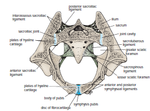
During pregnancy, the symphysis pubis and the ligaments of the sacroiliac and sacrococcygeal joints undergo softening in response to hormones, thus increasing the mobility and increasing the potential size of the pelvis during childbirth. The hormones responsible are estrogen and progesterone produced by the ovary and the placenta. An additional hormone, called relaxin, produced by these organs can also have a relaxing effect on the pelvic ligaments.

Changes with Age
Obliteration of the cavity in the sacroiliac joint occurs in both sexes after middle age.


Sacroiliac Joint Disease
The sacroiliac joint is innervated by the lower lumbar and sacral nerves so that disease in the joint can produce low back pain and pain referred along the sciatic nerve (sciatica). The sacroiliac joint is inaccessible to clinical examination. However, a small area located just medial to and below the posterior superior iliac spine is where the joint comes closest to the surface. In disease of the lumbosacral region, movements of the vertebral column in any direction cause pain in the lumbosacral part of the column. In sacroiliac disease, pain is extreme on rotation of the vertebral column and is worst at the end of forward flexion. The latter movement causes pain because the hamstring muscles (see page 465) hold the hip bones in position while the sacrum is rotating forward as the vertebral column is flexed


Fractures of the Pelvis-Fractures of the False Pelvis-Fractures of the True Pelvis-Fractures of the Sacrum and Coccyx-Minor Fractures of the Pelvis-Anatomy of Complications of Pelvic Fractures-

Fractures of the Pelvis

Fractures of the False Pelvis
direct trauma occasionally cause Fractures of the false pelvis. The upper part of the ilium is seldom displaced because of the attachment of the iliacus muscle on the inside and the gluteal muscles on the outside.

Fractures of the True Pelvis
The pelvis as a rigid ring not only as a basin. The ring is made up of the pubic rami, the ischium, the acetabulum, the ilium, and the sacrum, joined by strong ligaments at the sacroiliac and symphyseal joints. If the ring breaks at any one point, the fracture will be stable and no displacement will occur. However, if two breaks occur in the ring, the fracture will be unstable and displacement will occur, Fracture of bone on either side of the joint is more common than disruption of the joint.
because the postvertebral and abdominal muscles will shorten and elevate the lateral part of the pelvis. The break in the ring may occur not as the result of a fracture but as the result of disruption of the sacroiliac or symphyseal joints. The forces responsible for the disruption of the bony ring may be anteroposterior compression, lateral compression, or shearing.

A heavy fall on the greater trochanter of the femur may drive the head of the femur through the floor of the acetabulum into the pelvic cavity.

Fractures of the Sacrum and Coccyx
Fractures of the lateral mass of the sacrum may occur as part of a pelvic fracture. Fractures of the coccyx are rare. However, coccydynia is common and is usually caused by direct trauma to the coccyx, as in falling down a flight of concrete steps. The anterior surface of the coccyx can be palpated with a rectal examination.


Minor Fractures of the Pelvis
The anterior superior iliac spine may be pulled off by the forcible contraction of the sartorius muscle in athletes. In a similar manner, the anterior inferior iliac spine may be avulsed by the contraction of the rectus femoris muscle. The ischial tuberosity can be avulsed by the contraction of the hamstring muscles. Healing may occur by fibrous union, possibly resulting in elongation of the muscle unit and some reduction in muscular efficiency.


Anatomy of Complications of Pelvic Fractures
Fractures of the true pelvis are commonly associated with injuries to the soft pelvic tissues.
If damaged, the thin pelvic veins—namely, the internal iliac veins and their tributaries—that lie in the parietal pelvic fascia beneath the parietal peritoneum can be the source of a massive hemorrhage, which may be life threatening.
 The male urethra is often damaged, especially in vertical shear fractures that may disrupt the urogenital diaphragm.
The bladder, which lies immediately behind the pubis in both sexes, is occasionally damaged by spicules of bone; a full bladder is more likely to be injured than an empty bladder .
The rectum lies within the concavity of the sacrum and is protected and rarely damaged. Fractures of the sacrum or ischial spine may be thrust into the pelvic cavity, tearing the rectum. Nerve injuries can follow sacral fractures; the laying down of fibrous tissue around the anterior or posterior nerve roots or the branches of the sacral spinal nerves can result in persistent pain.

 
Damage to the sciatic nerve may occur in fractures involving the boundaries of the greater sciatic notch. The peroneal part of the sciatic nerve is most often involved, resulting in the inability of a conscious patient to dorsiflex the ankle joint or failure of an unconscious patient to reflexly plantar-flex (ankle jerk) the foot