Showing posts with label Minor. Show all posts

Sunday, 26 June 2016

The Axilla-Walls of the Axilla-Contents of the Axilla-Key Muscles in the Axilla-Pectoralis Minor-Clavipectoral Fascia-Absent Pectoralis Major-

The Axilla
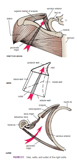
The axilla, or armpit, is a pyramid-shaped space between the upper part of the arm and the side of the chest. It forms an important passage for nerves, blood, and lymph vessels as they travel from the root of the neck to the upper limb. The upper end of the axilla, or apex, is directed into the root of the neck and is bounded in front by the clavicle, behind by the upper border of the scapula, and medially by the outer border of the first rib. The lower end, or base, is bounded in front by the anterior axillary fold (formed by the lower border of the pectoralis major muscle), behind by the posterior axillary fold (formed by the tendon of latissimus dorsi and the teres major muscle), and medially by the chest wall

Walls of the Axilla
The walls of the axilla are made up as follows:
■■ Anterior wall: By the pectoralis major, subclavius, and pectoralis minor muscles
■■ Posterior wall: By the subscapularis, latissimus dorsi, and teres major muscles from above down
■■ Medial wall: By the upper four or five ribs and the intercostal spaces covered by the serratus anterior muscle
■■ Lateral wall: By the coracobrachialis and biceps muscles in the bicipital groove of the humerus
The base is formed by the skin stretching between the anterior and posterior walls.

Contents of the Axilla
The axilla contains the axillary artery and its branches, which supply blood to the upper limb; the axillary vein and its tributaries, which drain blood from the upper limb; and lymph vessels and lymph nodes, which drain lymph from the upper limb and the breast and from the skin of the trunk, down as far as the level of the umbilicus. Lying among these structures in the axilla is an important nerve plexus, the brachial plexus, which innervates the upper limb. These structures are embedded in fat.


Key Muscles in the Axilla
Pectoralis Minor
The pectoralis minor is a thin triangular muscle that lies beneath the pectoralis major. It arises from the3rd, 4th, and 5th ribs and runs upward and laterally to be inserted by its apex into the coracoid process of the scapula. It crosses the axillary artery and the brachial plexus of nerves. It is used when describing the axillary artery to divide it into three parts
.
Clavipectoral Fascia
The clavipectoral fascia is a strong sheet of connective tissue that is attached above to the clavicle. Below, it splits to enclose the pectoralis minor muscle and then continues downward as the suspensory ligament of the axilla and joins the fascial floor of the armpit.

Absent Pectoralis Major
Occasionally, parts of the pectoralis major muscle may be absent. The sternocostal origin is the most commonly missing part, and this causes weakness in adduction and medial rotation of the shoulder joint.


Sunday, 19 June 2016

Fractures of the Pelvis-Fractures of the False Pelvis-Fractures of the True Pelvis-Fractures of the Sacrum and Coccyx-Minor Fractures of the Pelvis-Anatomy of Complications of Pelvic Fractures-

Fractures of the Pelvis

Fractures of the False Pelvis
direct trauma occasionally cause Fractures of the false pelvis. The upper part of the ilium is seldom displaced because of the attachment of the iliacus muscle on the inside and the gluteal muscles on the outside.

Fractures of the True Pelvis
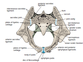
The pelvis as a rigid ring not only as a basin. The ring is made up of the pubic rami, the ischium, the acetabulum, the ilium, and the sacrum, joined by strong ligaments at the sacroiliac and symphyseal joints. If the ring breaks at any one point, the fracture will be stable and no displacement will occur. However, if two breaks occur in the ring, the fracture will be unstable and displacement will occur, Fracture of bone on either side of the joint is more common than disruption of the joint.
because the postvertebral and abdominal muscles will shorten and elevate the lateral part of the pelvis. The break in the ring may occur not as the result of a fracture but as the result of disruption of the sacroiliac or symphyseal joints. The forces responsible for the disruption of the bony ring may be anteroposterior compression, lateral compression, or shearing.

A heavy fall on the greater trochanter of the femur may drive the head of the femur through the floor of the acetabulum into the pelvic cavity.

Fractures of the Sacrum and Coccyx
Fractures of the lateral mass of the sacrum may occur as part of a pelvic fracture. Fractures of the coccyx are rare. However, coccydynia is common and is usually caused by direct trauma to the coccyx, as in falling down a flight of concrete steps. The anterior surface of the coccyx can be palpated with a rectal examination.


Minor Fractures of the Pelvis
The anterior superior iliac spine may be pulled off by the forcible contraction of the sartorius muscle in athletes. In a similar manner, the anterior inferior iliac spine may be avulsed by the contraction of the rectus femoris muscle. The ischial tuberosity can be avulsed by the contraction of the hamstring muscles. Healing may occur by fibrous union, possibly resulting in elongation of the muscle unit and some reduction in muscular efficiency.


Anatomy of Complications of Pelvic Fractures
Fractures of the true pelvis are commonly associated with injuries to the soft pelvic tissues.
If damaged, the thin pelvic veins—namely, the internal iliac veins and their tributaries—that lie in the parietal pelvic fascia beneath the parietal peritoneum can be the source of a massive hemorrhage, which may be life threatening.
 The male urethra is often damaged, especially in vertical shear fractures that may disrupt the urogenital diaphragm.
The bladder, which lies immediately behind the pubis in both sexes, is occasionally damaged by spicules of bone; a full bladder is more likely to be injured than an empty bladder .
The rectum lies within the concavity of the sacrum and is protected and rarely damaged. Fractures of the sacrum or ischial spine may be thrust into the pelvic cavity, tearing the rectum. Nerve injuries can follow sacral fractures; the laying down of fibrous tissue around the anterior or posterior nerve roots or the branches of the sacral spinal nerves can result in persistent pain.

 
Damage to the sciatic nerve may occur in fractures involving the boundaries of the greater sciatic notch. The peroneal part of the sciatic nerve is most often involved, resulting in the inability of a conscious patient to dorsiflex the ankle joint or failure of an unconscious patient to reflexly plantar-flex (ankle jerk) the foot