Showing posts with label Fractures. Show all posts

Wednesday, 6 July 2016

Fractures of the Femur-

Fractures of the Femur
Fractures of the neck of the femur are common and are of two types, subcapital and trochanteric. The subcapital fracture occurs in the elderly and is usually produced by a minor trip or stumble. Subcapital femoral neck fractures are particularly common in women after menopause. This gender predisposition is because of a thinning of the cortical and trabecular bone caused by estrogen deficiency. Avascular necrosis of the head is a common complication. If the fragments are not impacted, considerable displacement occurs. The strong muscles of the thigh, including the rectus femoris, the adductor muscles, and the hamstring muscles, pull the distal fragment upward, so that the leg is shortened (as measured from the anterior superior iliac spine to the adductor tubercle or medial malleolus). The gluteus maximus, the piriformis, the obturator internus, the gemelli, and the quadratus femoris rotate the distal fragment laterally, as seen by the toes pointing laterally.

Trochanteric fractures commonly occur in the young and middle aged as a result of direct trauma. The fracture line is extracapsular, and both fragments have a profuse blood supply. If the bone fragments are not impacted, the pull of the strong muscles will produce shortening and lateral rotation of the leg, as previously explained.

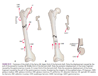
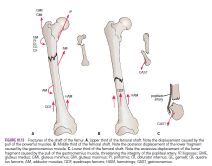
Fractures of the shaft of the femur usually occur in young and healthy persons. In fractures of the upper third of the shaft of the femur, the proximal fragment is flexed by the iliopsoas; abducted by the gluteus medius and minimus; and laterally rotated by the gluteus maximus, the piriformis, the obturator internus, the gemelli, and the quadratus femoris. The lower fragment is adducted by the adductor muscles, pulled upward by the hamstrings and quadriceps, and laterally rotated by the adductors and the weight of the foot.

In fractures of the middle third of the shaft of the femur, the distal fragment is pulled upward by the hamstrings and the quadriceps , resulting in considerable shortening. The distal fragment is also rotated backward by the pull of the two heads of the gastrocnemius. In fractures of the distal third of the shaft of the femur, the same displacement of the distal fragment occurs as seen in fractures of the middle third of the shaft. However, the distal fragment is smaller and is rotated backward by the gastrocnemius muscle to a greater degree and may exert pressure on the popliteal artery and interfere with the blood flow through the leg and foot.
From these accounts, it is clear that knowledge of the different actions of the muscles of the leg is necessary to understand the displacement of the fragments of a fractured femur.
Considerable traction on the distal fragment is usually required to overcome the powerful muscles and restore the limb to its correct length before manipulation and operative therapy to bring the proximal and distal fragments into correct alignment

Tuesday, 28 June 2016

Injuries to the Bones of the Hand-Dislocation of the lunate bone-Fractures of the metacarpal bones-Bennett’s fracture-Compartment Syndrome of the Forearm--Volkmann’s Ischemic Contracture

Injuries to the Bones of the Hand
Fracture of the scaphoid bone is common in young adults; unless treated effectively, the fragments will not unite, and permanent weakness and pain of the wrist will result, with the subsequent development of osteoarthritis. The fracture line usually goes through the narrowest part of the bone, which, because of its location, is bathed in synovial fluid. The blood vessels to the scaphoid enter its proximal and distal ends, although the blood supply is occasionally confined to its distal end. If the latter occurs, a fracture deprives the proximal fragment of its arterial supply, and this fragment undergoes avascular necrosis.
Deep tenderness in the anatomic snuffbox after a fall on the outstretched hand in a young adult makes one suspicious of a fractured scaphoid.

Dislocation of the lunate bone
occasionally occurs in young adults who fall on the outstretched hand in a way that causes hyperextension of the wrist joint. Involvement of the median nerve is common.

Fractures of the metacarpal bones
 can occur as a result of direct violence, such as the clenched fist striking a hard object.
The fracture always angulates dorsally. The “boxer’s fracture” commonly produces an oblique fracture of the neck of the fifth and sometimes the fourth metacarpal bones. The distal fragment is commonly displaced proximally, thus shortening the finger posteriorly.

Bennett’s fracture
is a fracture of the base of the metacarpal of the thumb caused when violence is applied along the long axis of the thumb or the thumb is forcefully abducted. The fracture is oblique and enters the carpometacarpal joint of the thumb, causing joint instability.
Fractures of the phalanges are common and usually follow direct injury.


Compartment Syndrome of the Forearm
The forearm is enclosed in a sheath of deep fascia, which is attached to the periosteum of the posterior subcutaneous border of the ulna. This fascial sheath, together with the interosseous membrane and fibrous intermuscular septa, divides the forearm into several compartments, each having its own muscles, nerves, and blood supply. There is very little room within each compartment, and any edema can cause secondary vascular compression of the blood vessels; the veins are first affected, and later the arteries.
Soft tissue injury is a common cause, and early diagnosis is critical. Early signs include altered skin sensation (caused by ischemia of the sensory nerves passing through the compartment),
pain disproportionate to any injury (caused by pressure on nerves within the compartment), pain on passive stretching of muscles that pass through the compartment (caused by muscle ischemia), tenderness of the skin over the compartment (a late sign caused by edema), and absence of capillary refill in the nail beds (caused by pressure on the arteries within the compartment). Once the diagnosis is made, the deep fascia must be incised surgically to decompress the affected compartment. A delay of as little as4 hours can cause irreversible damage to the muscles.


Volkmann’s Ischemic Contracture
Volkmann’s ischemic contracture is a contracture of the muscles of the forearm that commonly follows fractures of the distal end of the humerus or fractures of the radius and ulna. In this syndrome, a localized segment of the brachial artery goes into spasm, reducing the arterial flow to the flexor and the extensor muscles so that they undergo ischemic necrosis. The flexor muscles are larger than the extensor muscles, and they are therefore the ones mainly affected. The muscles are replaced by fibrous tissue, which contracts, producing the deformity. The arterial spasm is usually caused by an overtight cast, but in some cases the fracture itself may be responsible. The deformity can be explained only by understanding the anatomy of the region.

Three types of deformity exist:
■■ The long flexor muscles of the carpus and fingers are more contracted than the extensor muscles, and the wrist joint is flexed; the fingers are extended. If the wrist joint is extended passively, the fingers become flexed.
■■ The long extensor muscles to the fingers, which are inserted into the extensor expansion that is attached to the proximal phalanx, are greatly contracted; the metacarpophalangeal joints and the wrist joint are extended, and the interphalangeal joints of the fingers are flexed.
■■ Both the flexor and extensor muscles of the forearm are contracted. The wrist joint is flexed, the metacarpophalangeal joints are extended, and the interphalangeal joints are flexed.


-Fractures of the Radius and Ulna-Fractures of the head of the radius -Fractures of the neck of the radius-Fractures of the shafts of the radius-Fractures of the olecranon process-Olecranon Bursitis-The Metacarpals and Phalanges-

Fractures of the Radius and Ulna
Fractures of the head of the radius
can occur from falls on the outstretched hand. As the force is transmitted along the radius,
the head of the radius is driven sharply against the capitulum, splitting or splintering the head
.
Fractures of the neck of the radius
 occur in young children from falls on the outstretched hand.

Fractures of the shafts of the radius
 and ulna may or may not occur together. Displacement of the fragments is usually considerable and depends on the pull of the attached muscles. The proximal fragment of the radius is supinated by the supinator and the biceps brachii muscles. The distal fragment of the radius is pronated and pulled medially by the pronator quadratus muscle. The strength of the brachioradialis and extensor carpi radialis longus and brevis shortens and angulates the forearm. In fractures of the ulna, the ulna angulates posteriorly. To restore the normal movements of pronation and supination, the normal anatomic relationship of the radius, ulna, and interosseous membrane must be regained.
A fracture of one forearm bone may be associated with a dislocation of the other bone. In Monteggia’s fracture, for example, the shaft of the ulna is fractured by a force applied from behind.
There is a bowing forward of the ulnar shaft and an anterior dislocation of the radial head with rupture of the anular ligament. In Galeazzi’s fracture, the proximal third of the radius is fractured and the distal end of the ulna is dislocated at the distal radioulnar joint.

Fractures of the olecranon process
can result from a fall on the flexed elbow or from a direct blow. Depending on the location of the fracture line, the bony fragment may be displaced by the pull of the triceps muscle, which is inserted on the olecranon process. Avulsion fractures of part of the olecranon process can be produced by the pull of the triceps muscle. Good functional return after any of these fractures depends on the accurate anatomic reduction of the fragment.
Colles’ fracture is a fracture of the distal end of the radius resulting from a fall on the outstretched hand. It commonly occurs in patients older than 50 years. The force drives the distal fragment posteriorly and superiorly, and the distal articular
surface is inclined posteriorly. This posterior displacement produces a posterior bump, sometimes referred to as the “dinner-fork deformity” because the forearm and wrist resemble the shape of that eating utensil. Failure to restore the distal articular surface to its normal position will severely limit the range of flexion of the wrist joint.
Smith’s fracture is a fracture of the distal end of the radius and occurs from a fall on the back of the hand. It is a reversed Colles’ fracture because the distal fragment is displaced anteriorly

Olecranon Bursitis
A small subcutaneous bursa is present over the olecranon process of the ulna, and repeated trauma often produces chronic bursitis.

The Metacarpals and Phalanges
There are five metacarpal bones, each of which has a base, a shaft, and a head

The first metacarpal bone of the thumb is the shortest and most mobile. It does not lie in the same plane as the others but occupies a more anterior position. It is also rotated medially through a right angle so that its extensor surface is directed laterally and not backward.
The bases of the metacarpal bones articulate with the distal row of the carpal bones; the heads, which form the knuckles, articulate with the proximal phalanges.

 The shaft of each metacarpal bone is slightly concave forward and is triangular in transverse section. Its surfaces are posterior, lateral, and medial.
There are three phalanges for each of the fingers but only two for the thumb.


ش

Sunday, 26 June 2016

Humerus-Fractures of the Proximal End of the Humerus-Humeral Head Fractures-Greater Tuberosity Fractures-Lesser Tuberosity Fractures-Surgical Neck Fractures-Fractures of the Shaft of the Humerus-Fractures of the Distal End of the Humerus-

Humerus
The humerus articulates with the scapula at the shoulder joint and with the radius and ulna at the elbow joint. The upper end of the humerus has a head, which forms about one third of a sphere and articulates with the glenoid cavity of the scapula. Immediately below the head is the anatomic neck. Below the neck are the greater and lesser tuberosities, separated from each other by the bicipital groove. Where the upper end of the humerus joins the shaft is a narrow surgical neck. About halfway down the lateral aspect of the shaft is a roughened elevation called the deltoid tuberosity. Behind and below the tuberosity is a spiral groove, which accommodates the radial nerve The lower end of the humerus possesses the medial and lateral epicondyles for the attachment of muscles and ligaments, the rounded capitulum for articulation with the head of the radius, and the pulley-shaped trochlea for articulation with the trochlear notch of the ulna. Above the capitulum is the radial fossa, which receives the head of the radius when the elbow is flexed. Above the trochlea anteriorly is the coronoid fossa, which during the same movement receives the coronoid process of the ulna. Above the trochlea posteriorly is the olecranon fossa, which receives the olecranon process of the ulna when the elbow joint is extended

Fractures of the Proximal End of the Humerus
Humeral Head Fractures
Fractures of the humeral head can occur during the process of anterior and posterior dislocations of the shoulder joint. The fibrocartilaginous glenoid labrum of the scapula produces the fracture, and the labrum can become jammed in the defect, making reduction of the shoulder joint difficult
Greater Tuberosity Fractures
The greater tuberosity of the humerus can be fractured by direct trauma, displaced by the glenoid labrum during dislocation of the shoulder joint, or avulsed by violent contractions of the supraspinatus muscle. The bone fragment will have the attachments of the supraspinatus, teres minor, and infraspinatus muscles, whose tendons form part of the rotator cuff. When associated with a shoulder dislocation, severe tearing of the cuff with the fracture can result in the greater tuberosity remaining displaced posteriorly after the shoulder joint has been reduced. In this situation, open reduction of the fracture is necessary to attach the rotator cuff back into place.

Lesser Tuberosity Fractures
Occasionally, a lesser tuberosity fracture accompanies posterior dislocation of the shoulder joint. The bone fragment receives the insertion of the subscapularis tendon, a part of the rotator cuff.

Surgical Neck Fractures
The surgical neck of the humerus , which lies immediately distal to the lesser tuberosity, can be fractured by a direct blow on the lateral aspect of the shoulder or in an indirect manner by falling on the outstretched hand.

Fractures of the Shaft of the Humerus
Fractures of the humeral shaft are common; displacement of the fragments depends on the relation of the site of fracture to the insertion of the deltoid muscle. When the fracture line is proximal to the deltoid insertion, the proximal fragment is adducted by the pectoralis major, latissimus dorsi, and teres major muscles; the distal fragment is pulled proximally by the deltoid, biceps, and triceps. When the fracture is distal to the deltoid insertion, the proximal fragment is abducted by the deltoid, and the distal fragment is pulled proximally by the biceps and triceps. The radial nerve can be damaged where it lies in the spiral groove on the posterior surface of the humerus under cover of the triceps muscle.


Fractures of the Distal End of the Humerus
Supracondylar fractures are common in children and occur when the child falls on the outstretched hand with the elbow partially flexed. Injuries to the median, radial, and ulnar nerves are not uncommon, although function usually quickly returns after reduction of the fracture. Damage to or pressure on the brachial artery can occur at the time of the fracture or from swelling of the surrounding tissues; the circulation to the forearm may be interfered with, leading to Volkmann’s ischemic contractureThe medial epicondyle (Fig. 9.10) can be avulsed by themedial collateral ligament of the elbow joint if the forearm is forcibly abducted. The ulnar nerve can be injured at the time of the fracture, can become involved later in the repair process of the fracture (in the callus), or can undergo irritation
on the irregular bony surface after the bone fragments are
reunited
.


  .