Showing posts with label Pelvic. Show all posts

Tuesday, 21 June 2016

Uterine Tube-Uterine TubeFunction-Blood Supply-The Uterine Tube as a Conduit for Infection-Pelvic Inflammatory Disease-Ectopic Pregnancy- Tubal Ligation-

Uterine Tube
The two uterine tubes are each about 4 in. (10 cm) long and lie in the upper border of the broad ligament . Each connects the peritoneal cavity in the region of the ovary with the cavity of the uterus. The uterine tube is divided into four parts:
1. The infundibulum is the funnel-shaped lateral end that projects beyond the broad ligament and overlies the ovary. The free edge of the funnel has several fingerlike processes, known as fimbriae, which are draped over the ovary .
2. The ampulla is the widest part of the tube.
3. The isthmus is the narrowest part of the tube and lies just lateral to the uterus .
4. The intramural part is the segment that pierces the uterine wall .

Function
The uterine tube receives the ovum from the ovary and provides a site where fertilization of the ovum can take place (usually in the ampulla). It provides nourishment for the fertilized ovum and transports it to the cavity of the uterus. The tube serves as a conduit along which the spermatozoa travel to reach the ovum.

Blood Supply

Arteries
The uterine artery from the internal iliac artery and the ovarian artery from the abdominal aorta .

Veins
The veins correspond to the arteries.

Lymph Drainage
The internal iliac and para-aortic nodes.


Nerve Supply
Sympathetic and parasympathetic nerves from the inferior hypogastric plexuses

The Uterine Tube as a Conduit for Infection
The uterine tube lies in the upper free border of the broad ligament and is a direct route of communication from the vulva through the vagina and uterine cavity to the peritoneal cavity.

Pelvic Inflammatory Disease
The pathogenic organism(s) enter the body through sexual contact and ascend through the uterus and enter the uterine tubes. Salpingitis may follow, with leakage of pus into the peritoneal cavity, causing pelvic peritonitis. A pelvic abscess usually follows, or the infection spreads farther, causing general peritonitis.

Ectopic Pregnancy
Implantation and growth of a fertilized ovum may occur outside the uterine cavity in the wall of the uterine tube. This is a variety of ectopic pregnancy. There being no decidua formation in the tube, the eroding action of the trophoblast quickly destroys the wall of the tube. Tubal abortion or rupture of the tube, with the effusion of a large quantity of blood into the peritoneal cavity, is the common result.
The blood pours down into the rectouterine pouch (pouch of Douglas) or into the uterovesical pouch. The blood may quickly ascend into the general peritoneal cavity, giving rise to severe abdominal pain, tenderness, and guarding. Irritation of the subdiaphragmatic peritoneum (supplied by phrenic nerves C3, 4, and 5) may give rise to referred pain to the shoulder skin (supraclavicular nerves C3 and 4).


Tubal Ligation
Ligation and division of the uterine tubes is a method of obtaining permanent birth control and is usually restricted to women who already have children. The ova that are discharged from the ovarian follicles degenerate in the tube proximal to the obstruction. If, later, the woman wishes to have an additional child, restoration of the continuity of the uterine tubes can be attempted, and, in about 20% of women, fertilization occurs.




Sunday, 19 June 2016

Partial and Complete Prolapse of the Rectum-Cancer of the Rectum-Rectal Injuries-Pelvic Appendix

Partial and Complete Prolapse of the Rectum
Partial and complete prolapses of the rectum through the anus are relatively common clinical conditions. In partial prolapse, the rectal mucous membrane and submucous coat protrude for a short distance outside the anus. In complete prolapse, the whole thickness of the rectal wall protrudes through the anus. In both conditions, many causative factors may be involved. However, damage to the levatores ani muscles as the result of childbirth and poor muscle tone in the aged are important contributing factors. A complete rectal prolapse may be regarded as a sliding hernia through the pelvic diaphragm.

 
Cancer of the Rectum
Cancer of the rectum is a common clinical finding that remains localized to the rectal wall for a considerable time. At first, it tends to spread locally in the lymphatics around the circumference of the bowel. Later, it spreads upward and laterally along the lymph vessels, following the superior rectal and middle rectal arteries. Venous spread occurs late, and because the superior rectal vein is a tributary of the portal vein, the liver is a common site for secondary deposits.

Once the malignant tumor has extended beyond the confines of the rectal wall, knowledge of the anatomic relations of the rectum will enable a physician to assess the structures and organs likely to be involved. In both sexes, a posterior penetration involves the sacral plexus and can cause severe intractable pain down the leg in the distribution of the sciatic nerve. A lateral penetration may involve the ureter. An anterior penetration in the male may involve the prostate, seminal vesicles, or bladder; in the female, the vagina and uterus may be invaded. It is clear from the anatomic features of the rectum and its lymph drainage that a wide resection of the rectum with its lymphatic field offers the best chance of cure. When the tumor has spread to contiguous organs and is of a low grade of malignancy, some form of pelvic evisceration may be justifiable. It is most important for a medical student to remember that the interior of the lower part of the rectum can be examined by a gloved index finger introduced through the anal canal. The anal canal is about 1.5 in. (4 cm) long so that the pulp of the index finger can easily feel the mucous membrane lining the lower end of the rectum. Most cancers of the rectum can be diagnosed by this means. This examination can be extended in both sexes by placing the other hand on the lower part of the anterior abdominal wall. With the bladder empty, the anterior rectal wall can be examined bimanually. In the female, the placing of one finger in the vagina and another in the rectum may enable the physician to make a thorough examination of the lower part of the anterior rectal wall.


Rectal Injuries
The management of penetrating rectal injuries will be determined by the site of penetration relative to the peritoneal covering. The upper third of the rectum is covered on the anterior and lateral surfaces by peritoneum, the middle third is covered only on its anterior surface, and the lower third is devoid of a peritoneal covering. The treatment of penetration of the intraperitoneal portion of the rectum is identical to that of the colon, because the peritoneal cavity has been violated. In the case of penetration of the extraperitoneal portion, the rectum is treated by diverting the feces through a temporary abdominal colostomy, administering antibiotics, and repairing and draining the tissue in front of the sacrum.

Pelvic Appendix
If an inflamed appendix is hanging down into the pelvis, abdominal tenderness in the right iliac region may not be felt, but deep tenderness may be experienced above the symphysis pubis. Rectal examination (or vaginal examination in the female) may reveal tenderness of the peritoneum in the pelvis on the right side. If such an inflamed appendix perforates, a localized pelvic peritonitis may result.


Sex Differences of the Pelvis-Pelvic Joints Changes-Changes with Pregnancy-Changes with Age-Sacroiliac Joint Disease-

Sex Differences of the Pelvis
The sex differences of the bony pelvis are easily recognized.
The more obvious differences result from the adaptation of the female pelvis for childbearing. The stronger muscles in the male are responsible for the thicker bones and more prominent bony markings (Figs. 6.1 and 6.4).
■■ The false pelvis is shallow in the female and deep in the male.
■■ The pelvic inlet is transversely oval in the female but heart shaped in the male because of the indentation produced by the promontory of the sacrum in the male.
■■ The pelvic cavity is roomier in the female than in the male, and the distance between the inlet and the outlet is much shorter.
■■ The pelvic outlet is larger in the female than in the male.
 In the female the ischial tuberosities are everted and in the male they are turned in.
■■ The sacrum is shorter, wider, and flatter in the female than in the male.
■■ The subpubic angle, or pubic arch, is more rounded and wider in the female than in the male.
 
Pelvic Joints Changes
Changes with Pregnancy
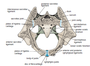
During pregnancy, the symphysis pubis and the ligaments of the sacroiliac and sacrococcygeal joints undergo softening in response to hormones, thus increasing the mobility and increasing the potential size of the pelvis during childbirth. The hormones responsible are estrogen and progesterone produced by the ovary and the placenta. An additional hormone, called relaxin, produced by these organs can also have a relaxing effect on the pelvic ligaments.

Changes with Age
Obliteration of the cavity in the sacroiliac joint occurs in both sexes after middle age.


Sacroiliac Joint Disease
The sacroiliac joint is innervated by the lower lumbar and sacral nerves so that disease in the joint can produce low back pain and pain referred along the sciatic nerve (sciatica). The sacroiliac joint is inaccessible to clinical examination. However, a small area located just medial to and below the posterior superior iliac spine is where the joint comes closest to the surface. In disease of the lumbosacral region, movements of the vertebral column in any direction cause pain in the lumbosacral part of the column. In sacroiliac disease, pain is extreme on rotation of the vertebral column and is worst at the end of forward flexion. The latter movement causes pain because the hamstring muscles (see page 465) hold the hip bones in position while the sacrum is rotating forward as the vertebral column is flexed


Fractures of the Pelvis-Fractures of the False Pelvis-Fractures of the True Pelvis-Fractures of the Sacrum and Coccyx-Minor Fractures of the Pelvis-Anatomy of Complications of Pelvic Fractures-

Fractures of the Pelvis

Fractures of the False Pelvis
direct trauma occasionally cause Fractures of the false pelvis. The upper part of the ilium is seldom displaced because of the attachment of the iliacus muscle on the inside and the gluteal muscles on the outside.

Fractures of the True Pelvis
The pelvis as a rigid ring not only as a basin. The ring is made up of the pubic rami, the ischium, the acetabulum, the ilium, and the sacrum, joined by strong ligaments at the sacroiliac and symphyseal joints. If the ring breaks at any one point, the fracture will be stable and no displacement will occur. However, if two breaks occur in the ring, the fracture will be unstable and displacement will occur, Fracture of bone on either side of the joint is more common than disruption of the joint.
because the postvertebral and abdominal muscles will shorten and elevate the lateral part of the pelvis. The break in the ring may occur not as the result of a fracture but as the result of disruption of the sacroiliac or symphyseal joints. The forces responsible for the disruption of the bony ring may be anteroposterior compression, lateral compression, or shearing.

A heavy fall on the greater trochanter of the femur may drive the head of the femur through the floor of the acetabulum into the pelvic cavity.

Fractures of the Sacrum and Coccyx
Fractures of the lateral mass of the sacrum may occur as part of a pelvic fracture. Fractures of the coccyx are rare. However, coccydynia is common and is usually caused by direct trauma to the coccyx, as in falling down a flight of concrete steps. The anterior surface of the coccyx can be palpated with a rectal examination.


Minor Fractures of the Pelvis
The anterior superior iliac spine may be pulled off by the forcible contraction of the sartorius muscle in athletes. In a similar manner, the anterior inferior iliac spine may be avulsed by the contraction of the rectus femoris muscle. The ischial tuberosity can be avulsed by the contraction of the hamstring muscles. Healing may occur by fibrous union, possibly resulting in elongation of the muscle unit and some reduction in muscular efficiency.


Anatomy of Complications of Pelvic Fractures
Fractures of the true pelvis are commonly associated with injuries to the soft pelvic tissues.
If damaged, the thin pelvic veins—namely, the internal iliac veins and their tributaries—that lie in the parietal pelvic fascia beneath the parietal peritoneum can be the source of a massive hemorrhage, which may be life threatening.
 The male urethra is often damaged, especially in vertical shear fractures that may disrupt the urogenital diaphragm.
The bladder, which lies immediately behind the pubis in both sexes, is occasionally damaged by spicules of bone; a full bladder is more likely to be injured than an empty bladder .
The rectum lies within the concavity of the sacrum and is protected and rarely damaged. Fractures of the sacrum or ischial spine may be thrust into the pelvic cavity, tearing the rectum. Nerve injuries can follow sacral fractures; the laying down of fibrous tissue around the anterior or posterior nerve roots or the branches of the sacral spinal nerves can result in persistent pain.

 
Damage to the sciatic nerve may occur in fractures involving the boundaries of the greater sciatic notch. The peroneal part of the sciatic nerve is most often involved, resulting in the inability of a conscious patient to dorsiflex the ankle joint or failure of an unconscious patient to reflexly plantar-flex (ankle jerk) the foot