Showing posts with label Radius. Show all posts

Wednesday, 29 June 2016

Development of the Upper Limb-Amelia-Congenital Absence of the Radius-Syndactyly-Lobster Hand-Brachydactyly-Floating Thumb-Polydactyly-Local Gigantism

Development of the Upper Limb
The limb buds appear during the sixth week of development as the result of a localized proliferation of somatopleuric mesenchyme. This causes the overlying ectoderm to bulge from the trunk as two pairs of flattened paddles. The arm buds develop before the leg buds and lie at the level of the lower six cervical and upper two thoracic segments. The flattened limb buds have a cephalic preaxial border and a caudal postaxial border. As the limb buds elongate, the anterior rami of the spinal nerves situated opposite the bases of the limb buds start to grow into the limbs.
The mesenchyme situated along the preaxial border becomes associated and innervated with the lower five cervical nerves, whereas the mesenchyme of the postaxial border becomes associated with the 8th cervical and 1st thoracic nerves.
Later, the mesenchymal masses divide into anterior and posterior groups, and the nerve trunks entering the base of each limb also divide into anterior and posterior divisions. The mesenchyme within the limbs differentiates into individual muscles that migrate within each limb. As a consequence of these two factors, the anterior rami of the spinal nerves become arranged in complicated plexuses that are found near the base of each limb so that the brachial plexus is formed.

Amelia
Absence of one or more limbs (amelia) or partial absence (ectromelia) may occur. A defective limb may possess a rudimentary hand at the extremity of the limb or a well-developed hand may spring from the shoulder with absence of the intermediate portion of the limb (phocomelia) .

Congenital Absence of the Radius
Occasionally, the radius is congenitally absent and the growth of the ulna pushes the hand laterally.

Syndactyly
In syndactyly, there is webbing of the fingers. It is usually bilateral and often familial. Plastic repair of the fingers is carried out at the age of 5 years.


Lobster Hand
Lobster hand is a form of syndactyly that is associated with a central cleft dividing the hand into two parts. It is a heredofamilial disorder, for which plastic surgery is indicated where possible.

Brachydactyly
In brachydactyly, there is an absence of one or more phalanges in several fingers. Provided that the thumb is functioning normally, surgery is not indicated .

Floating Thumb
A floating thumb results if the metacarpal bone of the thumb is absent but the phalanges are present. Plastic surgery is indicated where possible to improve the functional capabilities of the hand.

Polydactyly
In polydactyly, one or more extra digits develop. It tends to run in families. The additional digits are removed surgically.

Local Gigantism
Macrodactyly affects one or more digits; these may be of adult size at birth, but the size usually diminishes with age. Surgical removal may be necessary.


Tuesday, 28 June 2016

-Fractures of the Radius and Ulna-Fractures of the head of the radius -Fractures of the neck of the radius-Fractures of the shafts of the radius-Fractures of the olecranon process-Olecranon Bursitis-The Metacarpals and Phalanges-

Fractures of the Radius and Ulna
Fractures of the head of the radius
can occur from falls on the outstretched hand. As the force is transmitted along the radius,
the head of the radius is driven sharply against the capitulum, splitting or splintering the head
.
Fractures of the neck of the radius
 occur in young children from falls on the outstretched hand.

Fractures of the shafts of the radius
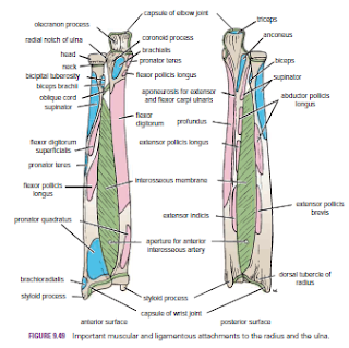
 and ulna may or may not occur together. Displacement of the fragments is usually considerable and depends on the pull of the attached muscles. The proximal fragment of the radius is supinated by the supinator and the biceps brachii muscles. The distal fragment of the radius is pronated and pulled medially by the pronator quadratus muscle. The strength of the brachioradialis and extensor carpi radialis longus and brevis shortens and angulates the forearm. In fractures of the ulna, the ulna angulates posteriorly. To restore the normal movements of pronation and supination, the normal anatomic relationship of the radius, ulna, and interosseous membrane must be regained.
A fracture of one forearm bone may be associated with a dislocation of the other bone. In Monteggia’s fracture, for example, the shaft of the ulna is fractured by a force applied from behind.
There is a bowing forward of the ulnar shaft and an anterior dislocation of the radial head with rupture of the anular ligament. In Galeazzi’s fracture, the proximal third of the radius is fractured and the distal end of the ulna is dislocated at the distal radioulnar joint.

Fractures of the olecranon process
can result from a fall on the flexed elbow or from a direct blow. Depending on the location of the fracture line, the bony fragment may be displaced by the pull of the triceps muscle, which is inserted on the olecranon process. Avulsion fractures of part of the olecranon process can be produced by the pull of the triceps muscle. Good functional return after any of these fractures depends on the accurate anatomic reduction of the fragment.
Colles’ fracture is a fracture of the distal end of the radius resulting from a fall on the outstretched hand. It commonly occurs in patients older than 50 years. The force drives the distal fragment posteriorly and superiorly, and the distal articular
surface is inclined posteriorly. This posterior displacement produces a posterior bump, sometimes referred to as the “dinner-fork deformity” because the forearm and wrist resemble the shape of that eating utensil. Failure to restore the distal articular surface to its normal position will severely limit the range of flexion of the wrist joint.
Smith’s fracture is a fracture of the distal end of the radius and occurs from a fall on the back of the hand. It is a reversed Colles’ fracture because the distal fragment is displaced anteriorly

Olecranon Bursitis
A small subcutaneous bursa is present over the olecranon process of the ulna, and repeated trauma often produces chronic bursitis.

The Metacarpals and Phalanges
There are five metacarpal bones, each of which has a base, a shaft, and a head

The first metacarpal bone of the thumb is the shortest and most mobile. It does not lie in the same plane as the others but occupies a more anterior position. It is also rotated medially through a right angle so that its extensor surface is directed laterally and not backward.
The bases of the metacarpal bones articulate with the distal row of the carpal bones; the heads, which form the knuckles, articulate with the proximal phalanges.

 The shaft of each metacarpal bone is slightly concave forward and is triangular in transverse section. Its surfaces are posterior, lateral, and medial.
There are three phalanges for each of the fingers but only two for the thumb.


ش