Showing posts with label Head. Show all posts

Tuesday, 28 June 2016

-Fractures of the Radius and Ulna-Fractures of the head of the radius -Fractures of the neck of the radius-Fractures of the shafts of the radius-Fractures of the olecranon process-Olecranon Bursitis-The Metacarpals and Phalanges-

Fractures of the Radius and Ulna
Fractures of the head of the radius
can occur from falls on the outstretched hand. As the force is transmitted along the radius,
the head of the radius is driven sharply against the capitulum, splitting or splintering the head
.
Fractures of the neck of the radius
 occur in young children from falls on the outstretched hand.

Fractures of the shafts of the radius
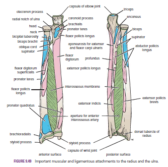
 and ulna may or may not occur together. Displacement of the fragments is usually considerable and depends on the pull of the attached muscles. The proximal fragment of the radius is supinated by the supinator and the biceps brachii muscles. The distal fragment of the radius is pronated and pulled medially by the pronator quadratus muscle. The strength of the brachioradialis and extensor carpi radialis longus and brevis shortens and angulates the forearm. In fractures of the ulna, the ulna angulates posteriorly. To restore the normal movements of pronation and supination, the normal anatomic relationship of the radius, ulna, and interosseous membrane must be regained.
A fracture of one forearm bone may be associated with a dislocation of the other bone. In Monteggia’s fracture, for example, the shaft of the ulna is fractured by a force applied from behind.
There is a bowing forward of the ulnar shaft and an anterior dislocation of the radial head with rupture of the anular ligament. In Galeazzi’s fracture, the proximal third of the radius is fractured and the distal end of the ulna is dislocated at the distal radioulnar joint.

Fractures of the olecranon process
can result from a fall on the flexed elbow or from a direct blow. Depending on the location of the fracture line, the bony fragment may be displaced by the pull of the triceps muscle, which is inserted on the olecranon process. Avulsion fractures of part of the olecranon process can be produced by the pull of the triceps muscle. Good functional return after any of these fractures depends on the accurate anatomic reduction of the fragment.
Colles’ fracture is a fracture of the distal end of the radius resulting from a fall on the outstretched hand. It commonly occurs in patients older than 50 years. The force drives the distal fragment posteriorly and superiorly, and the distal articular
surface is inclined posteriorly. This posterior displacement produces a posterior bump, sometimes referred to as the “dinner-fork deformity” because the forearm and wrist resemble the shape of that eating utensil. Failure to restore the distal articular surface to its normal position will severely limit the range of flexion of the wrist joint.
Smith’s fracture is a fracture of the distal end of the radius and occurs from a fall on the back of the hand. It is a reversed Colles’ fracture because the distal fragment is displaced anteriorly
Olecranon Bursitis
A small subcutaneous bursa is present over the olecranon process of the ulna, and repeated trauma often produces chronic bursitis.

The Metacarpals and Phalanges
There are five metacarpal bones, each of which has a base, a shaft, and a head

The first metacarpal bone of the thumb is the shortest and most mobile. It does not lie in the same plane as the others but occupies a more anterior position. It is also rotated medially through a right angle so that its extensor surface is directed laterally and not backward.
The bases of the metacarpal bones articulate with the distal row of the carpal bones; the heads, which form the knuckles, articulate with the proximal phalanges.

 The shaft of each metacarpal bone is slightly concave forward and is triangular in transverse section. Its surfaces are posterior, lateral, and medial.
There are three phalanges for each of the fingers but only two for the thumb.


ش

Sunday, 26 June 2016

Humerus-Fractures of the Proximal End of the Humerus-Humeral Head Fractures-Greater Tuberosity Fractures-Lesser Tuberosity Fractures-Surgical Neck Fractures-Fractures of the Shaft of the Humerus-Fractures of the Distal End of the Humerus-

Humerus
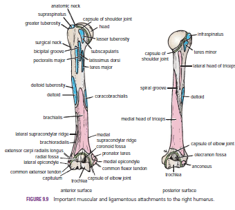
The humerus articulates with the scapula at the shoulder joint and with the radius and ulna at the elbow joint. The upper end of the humerus has a head, which forms about one third of a sphere and articulates with the glenoid cavity of the scapula. Immediately below the head is the anatomic neck. Below the neck are the greater and lesser tuberosities, separated from each other by the bicipital groove. Where the upper end of the humerus joins the shaft is a narrow surgical neck. About halfway down the lateral aspect of the shaft is a roughened elevation called the deltoid tuberosity. Behind and below the tuberosity is a spiral groove, which accommodates the radial nerve The lower end of the humerus possesses the medial and lateral epicondyles for the attachment of muscles and ligaments, the rounded capitulum for articulation with the head of the radius, and the pulley-shaped trochlea for articulation with the trochlear notch of the ulna. Above the capitulum is the radial fossa, which receives the head of the radius when the elbow is flexed. Above the trochlea anteriorly is the coronoid fossa, which during the same movement receives the coronoid process of the ulna. Above the trochlea posteriorly is the olecranon fossa, which receives the olecranon process of the ulna when the elbow joint is extended

Fractures of the Proximal End of the Humerus
Humeral Head Fractures
Fractures of the humeral head can occur during the process of anterior and posterior dislocations of the shoulder joint. The fibrocartilaginous glenoid labrum of the scapula produces the fracture, and the labrum can become jammed in the defect, making reduction of the shoulder joint difficult
Greater Tuberosity Fractures
The greater tuberosity of the humerus can be fractured by direct trauma, displaced by the glenoid labrum during dislocation of the shoulder joint, or avulsed by violent contractions of the supraspinatus muscle. The bone fragment will have the attachments of the supraspinatus, teres minor, and infraspinatus muscles, whose tendons form part of the rotator cuff. When associated with a shoulder dislocation, severe tearing of the cuff with the fracture can result in the greater tuberosity remaining displaced posteriorly after the shoulder joint has been reduced. In this situation, open reduction of the fracture is necessary to attach the rotator cuff back into place.

Lesser Tuberosity Fractures
Occasionally, a lesser tuberosity fracture accompanies posterior dislocation of the shoulder joint. The bone fragment receives the insertion of the subscapularis tendon, a part of the rotator cuff.

Surgical Neck Fractures
The surgical neck of the humerus , which lies immediately distal to the lesser tuberosity, can be fractured by a direct blow on the lateral aspect of the shoulder or in an indirect manner by falling on the outstretched hand.

Fractures of the Shaft of the Humerus
Fractures of the humeral shaft are common; displacement of the fragments depends on the relation of the site of fracture to the insertion of the deltoid muscle. When the fracture line is proximal to the deltoid insertion, the proximal fragment is adducted by the pectoralis major, latissimus dorsi, and teres major muscles; the distal fragment is pulled proximally by the deltoid, biceps, and triceps. When the fracture is distal to the deltoid insertion, the proximal fragment is abducted by the deltoid, and the distal fragment is pulled proximally by the biceps and triceps. The radial nerve can be damaged where it lies in the spiral groove on the posterior surface of the humerus under cover of the triceps muscle.


Fractures of the Distal End of the Humerus
Supracondylar fractures are common in children and occur when the child falls on the outstretched hand with the elbow partially flexed. Injuries to the median, radial, and ulnar nerves are not uncommon, although function usually quickly returns after reduction of the fracture. Damage to or pressure on the brachial artery can occur at the time of the fracture or from swelling of the surrounding tissues; the circulation to the forearm may be interfered with, leading to Volkmann’s ischemic contractureThe medial epicondyle (Fig. 9.10) can be avulsed by themedial collateral ligament of the elbow joint if the forearm is forcibly abducted. The ulnar nerve can be injured at the time of the fracture, can become involved later in the repair process of the fracture (in the callus), or can undergo irritation
on the irregular bony surface after the bone fragments are
reunited
.


  .

Sunday, 19 June 2016

Sacral Plexus-Pressure from the Fetal Head-Invasion by Malignant Tumors-Referred Pain from the Obturator Nerve-Caudal Anesthesia (Analgesia)

Sacral Plexus
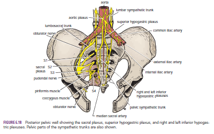
The sacral plexus lies in front of the piriformis muscle on the posterior pelvic wall. It is formed from the anterior rami of the 4th and 5th lumbar nerves and the anterior rami of the first, second, third, and fourth sacral nerves. The fourth lumbar nerve joins the fifth lumbar nerve to form the lumbosacral trunk. The lumbosacral trunk passes down into the pelvis and joins the sacral nerves as they emerge from the anterior sacral foramina.

 
Pressure from the Fetal Head
when the fetal head has descended into the pelvis During the later stages of pregnancy, , the mother often complains of discomfort or aching pain extending down one of the lower limbs. The discomfort, caused by pressure from the fetal head, is often relieved by changing position, such as lying on the side in bed.

Invasion by Malignant Tumors
The nerves of the sacral plexus can become invaded by malignant tumors extending from neighboring viscera. A carcinoma of the rectum, for example, can cause severe intractable pain down the lower limbs.

Referred Pain from the Obturator Nerve
The obturator nerve lies on the lateral wall of the pelvis and supplies the parietal peritoneum. An inflamed appendix hanging down into the pelvic cavity could cause irritation of the obturator nerve endings, leading to referred pain down the inner side of the right thigh. Inflammation of the ovaries can produce similar symptoms.

Caudal Anesthesia (Analgesia)
Anesthetic solutions can be injected into the sacral canal through the sacral hiatus. The solutions then act on the spinal roots of the 2nd, 3rd, 4th and 5th sacral and coccygeal segments of the cord as they emerge from the dura mater. The roots of higher spinal segments can also be blocked by this method. The needle must be confined to the lower part of the sacral canal, because the meninges extend down as far as the lower border of the second sacral vertebra. Caudal anesthesia is used in obstetrics to block pain fibers from the cervix of the uterus and to anesthetize the perineum


Pancreas-Relations-Trauma of the Pancreas-Cancer of the Head of the Pancreas and the Bile Duct-The Pancreatic Tail and Splenectomy-Diagnosis of Pancreatic Disease

Pancreas
The pancreas is both an exocrine and endocrine gland.
 The exocrine portion of the gland function is  producing secretion that contains enzymes capable of hydrolyzing proteins, fats, and carbohydrates. islets of Langerhans  of the gland, the pancreatic islets (The endocrine portion), produces the hormones insulin and glucagon, which play a key role in carbohydrate metabolism.
It is is an elongated structure that lies in the epigastrium and the left upper quadrant. It is soft and lobulated and situated on the posterior abdominal wall behind the peritoneum. It crosses the transpyloric plane. The pancreas is divided into a head, neck, body, and tail.

The head of the pancreas is disc shaped and lies within the concavity of the duodenum. the uncinate process,is  A part of the head extends to the left behind the superior mesenteric vessels .
The neck which connects the head to the body is the constricted portion of the pancreas and. It lies in front of the beginning of the portal vein and the origin of the superior mesenteric artery from the aorta.
The body runs upward and to the left across the midline . It is somewhat triangular in cross section. The tail passes forward in the splenicorenal ligament and comes in contact with the hilum of the spleen

Relations
■■ Anteriorly: From right to left: the transverse colon and the attachment of the transverse mesocolon, the lesser sac, and the stomach
■■ Posteriorly: From right to left: the bile duct, the portal and splenic veins, the inferior vena cava, the aorta, the origin of the superior mesenteric artery, the left psoas muscle, the left suprarenal gland, the left kidney, and the hilum of the spleen

Trauma of the Pancreas
The pancreas is is well protected because it is deeply placed within the abdomen and  protected by the costal margin and the anterior abdominal wall. However, blunt trauma, such as in a sports injury when a sudden blow to the abdomen occurs, can compress and tear the pancreas against the vertebral column. The pancreas is most commonly damaged by gunshot or stab wounds. Damaged pancreatic tissue releases activated pancreatic enzymes that produce the signs and symptoms of acute peritonitis.
Cancer of the Head of the Pancreas and the Bile Duct
cancer of the head of the pancreas often causes obstructive jaundice Because of the close relation of the head of the pancreas to the bile duct,.

The Pancreatic Tail and Splenectomy
The presence of the tail of the pancreas in the splenicorenal ligament sometimes results in its damage during splenectomy. The damaged pancreas releases enzymes that start to digest surrounding tissues, with serious consequences

Diagnosis of Pancreatic Disease
The deep location of the pancreas sometimes gives rise to problems of diagnosis for the following reasons:
■■ Pain from the pancreas is commonly referred to the back.
■■ Because the pancreas lies behind the stomach and transverse colon, disease of the gland can be confused with that of the stomach or transverse colon.
■■ Inflammation of the pancreas can spread to the peritoneum forming the posterior wall of the lesser sac. This in turn can lead to adhesions and the closing off of the lesser sac to form a pseudocyst.