Showing posts with label arteries. Show all posts

Sunday, 28 August 2016

Arteries of the Gluteal Region-Superior Gluteal Artery-Inferior Gluteal Artery-The- Trochanteric Anastomosis

Arteries of the Gluteal Region
Superior Gluteal Artery
The superior gluteal artery is a branch from the internal iliac artery and enters the gluteal region through the upper part of the greater sciatic foramen above the piriformis. It divides into branches that are distributed throughout the gluteal region.

 
Inferior Gluteal Artery
The inferior gluteal artery is a branch of the internal iliac artery and enters the gluteal region through the lower part of the greater sciatic foramen, below the piriformis. It divides into numerous branches that are distributed throughout the gluteal region.

The Trochanteric Anastomosis
The trochanteric anastomosis provides the main blood supply to the head of the femur. The nutrient arteries pass along the femoral neck beneath the capsule. The following arteries take part in the anastomosis: the superior gluteal artery, the inferior gluteal artery, the medial femoral circumflex artery, and the lateral femoral circumflex artery.

The Cruciate Anastomosis
The cruciate anastomosis is situated at the level of the lesser trochanter of the femur and, together with the trochanteric anastomosis, provides a connection between the internal iliac and the femoral arteries. The following arteries take part in the anastomosis: the inferior gluteal artery, the medial femoral circumflex artery, the lateral femoral circumflex artery, and the first perforating artery, a branch of the profunda artery.

Veins of the Lower Limb
The veins of the lower limb can be divided into three groups: superficial, deep, and perforating. The superficial veins consist of the great and small saphenous veins and their tributaries, which are situated beneath the skin in the superficial fascia.
The constant position of the great saphenous vein in front of the medial malleolus should be remembered for patients requiring emergency blood transfusion. The deep veins are the venae comitantes to the anterior and posterior tibial arteries, the popliteal vein, and the femoral veins and their tributaries. The perforating veins are communicating vessels that run between the superficial and deep veins. Many of these veins are found particularly in the region of the ankle and the medial side of the lower part of the leg. They possess valves that are arranged to prevent the flow of blood from the deep to the superficial veins.


Wednesday, 29 June 2016

Arterial Injury of the upper limb-Palpation and Compression of Arteries-Allen Test-Arterial Innervation and Raynaud’s Disease-

Arterial Injury of the upper limb
The arteries of the upper limb can be damaged by penetrating wounds or may require ligation in amputation operations.
Because of the existence of an adequate collateral circulation around the shoulder, elbow, and wrist joints, ligation of the main arteries of the upper limb is not followed by tissue necrosis or gangrene, provided, of course, that the arteries forming the collateral circulation are not diseased and the patient’s general circulation is satisfactory. Nevertheless, it can take days or weeks for the collateral vessels to open sufficiently to provide the distal part of the limb with the same volume of blood as previously supplied by the main artery.

Palpation and Compression of Arteries
A clinician must know where the arteries of the upper limb can be palpated or compressed in an emergency. The subclavian artery, as it crosses the first rib to become the axillary artery, can be palpated in the root of the posterior triangle of the neck. The artery can be compressed here against the first rib to stop a catastrophic hemorrhage. The third part of the axillary artery can be felt in the axilla as it lies in front of the teres major muscle. The brachial artery can be palpated in the arm as it lies on the brachialis and is overlapped from the lateral side by the biceps brachii.
The radial artery lies superficially in front of the distal end of the radius, between the tendons of the brachioradialis and flexor carpi radialis; it is here that the clinician takes the radial pulse. If the pulse cannot be felt, try feeling for the radial artery on the other wrist; occasionally, a congenitally abnormal radial artery can be difficult to feel. The radial artery can be less easily felt as it crosses the anatomic snuffbox.
The ulnar artery can be palpated as it crosses anterior to the flexor retinaculum in company with the ulnar nerve. The artery lies lateral to the pisiform bone, separated from it by the ulnar nerve. The artery is commonly damaged here in laceration wounds in front of the wrist.

Allen Test
The Allen test is used to determine the patency of the ulnar and radial arteries. With the patient’s hands resting in the lap, compress the radial arteries against the anterior surface of each radius and ask the patient to tightly clench the fists. The clenching of the fists closes off the superficial and deep palmar arterial arches. When the patient is asked to open the hands, the skin of the palms is at first white, and then normally the blood quickly flows into the arches through the ulnar arteries, causing the palms to promptly turn pink. This establishes that the ulnar arteries are patent. The patency of the radial arteries can be established by repeating the test but this time compressing the ulnar arteries as they lie lateral to the pisiform bones.

Arterial Innervation and Raynaud’s Disease
The arteries of the upper limb are innervated by sympathetic nerves. The preganglionic fibers originate from cell bodies in the 2nd to 8th thoracic segments of the spinal cord. They ascend in the sympathetic trunk and synapse in the middle cervical, inferior cervical, 1st thoracic, or stellate ganglia. The postganglionic fibers join the nerves that form the brachial plexus and are distributed to the arteries within the branches of the plexus.
For example, the digital arteries of the fingers are supplied by postganglionic sympathetic fibers that run in the digital nerves. Vasospastic diseases involving digital arterioles, such as Raynaud’s disease, may require a cervicodorsal preganglionic sympathectomy to prevent necrosis of the fingers. The operation is followed by arterial vasodilatation, with consequent increased blood flow to the upper limb.


Sunday, 26 June 2016

The Breasts-The Breasts at Puberty-Young Women-The Breast atPregnancy-Postmenopause-Blood Supply to Breast-Arteries-Veins-Lymph Drainage-

The Breasts
The breasts, they are situated in the pectoral region so they are not anatomically part of the upper limb and their blood supply and lymphatic drainage is largely into the armpit. Their clinical importance cannot be overemphasized.
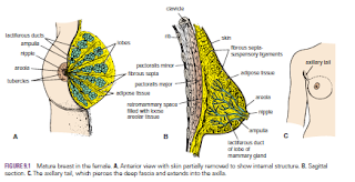
The breasts are specialized accessory glands of the skin that secrete milk. They are present in both sexes. In males and immature females, they are similar in structure. The nipples are small and surrounded by a colored area of skin called the areola. The breast tissue consists of a system of ducts embedded in connective tissue that does not extend beyond the margin of the areola.

Puberty
At puberty in females, the breasts gradually enlarge and assume their hemispherical shape under the influence of the ovarian hormones. The ducts elongate, but the increased size of the glands is mainly from the deposition of fat. The base of the breast extends from the 2nd to 6th rib and from the lateral margin of the sternum to the midaxillary line. The greater part of the gland lies in the superficial fascia. A small part, called the axillary tail, extends upward and laterally, pierces the deep fascia at the lower border of the pectoralis major muscle, and enters the axilla.
Each breast consists of 15 to 20 lobes, which radiate out from the nipple. The main duct from each lobe opens separately on the summit of the nipple and possesses a dilated ampulla just before its termination. The base of the nipple is surrounded by the areola. Tiny tubercles on the areola are produced by the underlying areolar glands.
The lobes of the gland are separated by fibrous septa that serve as suspensory ligaments. Behind the breasts is a space filled by loose connective tissue called the retromammary space.

Young Women
In young women, the breasts tend to protrude forward from a circular base.


Pregnancy
Early In the early months of pregnancy, there is a rapid increase in length and branching in the duct system. The secretory alveoli develop at the ends of the smaller ducts, and the connective tissue becomes filled with expanding and budding secretory alveoli. The vascularity of the connective tissue also increases to provide adequate nourishment for the developing gland. The nipple enlarges, and the areola becomes darker and more extensive as a result of increased deposits of melanin pigment in the epidermis. The areolar glands enlarge and become more active.
Late During the second half of pregnancy, the growth process slows. The breasts, however, continue to enlarge, mostly because of the distention of the secretory alveoli with the fluid secretion called colostrum. Postweaning Once the baby has been weaned, the breasts return to their inactive state. The remaining milk is absorbed, the secretory alveoli shrink, and most of them disappear. The interlobular connective tissue thickens. The breasts and the nipples shrink and return nearly to their original size. The pigmentation of the areola fades, but the area never lightens to its original color.

Postmenopause
After the menopause, the breast atrophies. Most of the secretory alveoli disappear, leaving behind the ducts. The amount of adipose tissue may increase or decrease. The breasts tend to shrink in size and become more pendulous. The atrophy after menopause is caused by the absence of ovarian estrogens and progesterone

Blood Supply

Arteries
The branches to the breasts include the perforating branches of the internal thoracic artery and the intercostal arteries. The axillary artery also supplies the gland via its lateral thoracic and thoracoacromial branches.

Veins
The veins correspond to the arteries.


Lymph Drainage
The lymph drainage of the mammary gland is of great clinical importance because of the frequent development of cancer in the gland and the subsequent dissemination of the malignant cells along the lymph vessels to the lymph nodes.
The lateral quadrants of the breast drain into the anterior axillary or pectoral group of nodes (situated just posterior to the lower border of the pectoralis major muscle). The medial quadrants drain by means of vessels that pierce the intercostal spaces and enter the internal thoracic group of nodes (situated within the thoracic cavity along the course of the internal thoracic artery). A few lymph vessels follow the posterior intercostal arteries and drain posteriorly into the posterior intercostal nodes (situated along the course of the posterior intercostal arteries); some vessels communicate with the lymph vessels of the opposite breast and with those of the anterior abdominal wall.


Sunday, 19 June 2016

Trauma to the Abdominal Aorta-Obliteration of the Abdominal Aorta and Iliac Arteries-Trauma to the Inferior Vena Cava- Compression of the Inferior Vena Cava-

Trauma to the Abdominal Aorta
Blunt trauma to the aorta is most commonly caused by headon automobile crashes. Rupture of the tunica intima and media occurs and is quickly followed by rupture of the turnica adventitia. The initial rupture of the intima and media is probably mainly caused by the sudden compression of the aorta against the vertebral column, while the delayed rupture of the adventitia is caused by the aortic blood pressure. Unless quickly diagnosed by MRI, and surgical treatment instituted, death follows.

 
Obliteration of the Abdominal Aorta and Iliac Arteries
Gradual occlusion of the bifurcation of the abdominal aorta, produced by atherosclerosis, results in the characteristic clinical symptoms of pain in the legs on walking (claudication) and impotence, the latter caused by lack of blood in the internal iliac arteries. In otherwise healthy individuals, surgical treatment by thromboendarterectomy or a bypass graft should be considered. Because the progress of the disease is slow, some collateral circulation is established, but it is physiologically inadequate. However, the collateral blood flow does prevent tissue death in both lower limbs, although skin ulcers may occur.

Trauma to the Inferior Vena Cava
Injuries to the inferior vena cava are commonly lethal, despite the fact that the contained blood is under low pressure. The anatomic inaccessibility of the vessel behind the liver, duodenum, and mesentery of the small intestine and the blocking presence of the right costal margin make a surgical approach difficult. Moreover, the thin wall of the vena cava makes it prone to extensive tears.
Because of the multiple anastomoses of the tributaries of the inferior vena cava, it is impossible in an emergency to ligate the vessel. Most patients have venous congestion of the lower limbs.

Compression of the Inferior Vena Cava
the enlarged uterus during the later stages of pregnancy is commonly compress The inferior vena cava. This produces edema of the ankles and feet and temporary varicose veins. Malignant retroperitoneal tumors can cause severe compression and eventual blockage of the inferior vena cava. This results in the dilatation of the extensive anastomoses of the tributaries. This alternative pathway for the blood to return to the right atrium of the heart is commonly referred to as the caval–caval shunt. The same pathway comes into effect in patients with a superior mediastinal tumor compressing the superior vena cava. Clinically, the enlarged subcutaneous anastomosis between the lateral thoracic vein, a tributary of the axillary vein; and the superficial epigastric vein, a tributary of the femoral vein, may be seen on the thoracoabdominal wall