Showing posts with label Drainage. Show all posts

Sunday, 26 June 2016

Breast Examination-Mammography-Breast Abscess-Lymph Drainage and Carcinoma of the Breast-Carcinoma in the Male Breast-

Breast Examination
 Because The breast is one of the common sites of cancer in women. It is also the site of different types of benign tumors and may be subject to acute inflammation and abscess formation. For these reasons, the clinical personnel must be familiar with the development, structure, and lymph drainage of this organ.
With the patient undressed to the waist and sitting upright, the breasts are first inspected for symmetry. Some degree of asymmetry is common and is the result of unequal breast development. Any swelling should be noted. A swelling can be caused by an underlying tumor, a cyst, or abscess formation. The nipples should be carefully examined for evidence of retraction. A carcinoma within the breast substance can cause retraction of the nipple by pulling on the lactiferous ducts. The patient is then asked to lie down so that the breasts can be palpated against the underlying thoracic wall. Finally, the patient is asked to sit up again and raise both arms above her head. With this maneuver, a carcinoma tethered to the skin, the suspensory ligaments, or the lactiferous ducts produces dimpling of the skin or retraction of the nipple.

Mammography
Mammography is a radiographic examination of the breast. This technique is extensively used for screening the breasts for benign and malignant tumors and cysts. Extremely low doses of x-rays are used so that the dangers are minimal, and the examination can be repeated often. Its success is based on the fact that a lesion measuring only a few millimeters in diameter can be detected long before it is felt by clinical examination.

Breast Abscess
during lactation An acute infection of the mammary gland may occur. Pathogenic bacteria gain entrance to the breast tissue through a crack in the nipple. Because of the presence of the fibrous septa, the infection remains localized to one compartment or lobe to begin with. Abscesses should be drained through a radial incision to avoid spreading of the infection into neighboring compartments; a radial incision also minimizes the damage to the radially arranged ducts.


Lymph Drainage and Carcinoma of the Breast
The importance of knowing the lymph drainage of the breast in relation to the spread of cancer from that organ cannot be overemphasized. The lymph vessels from the medial quadrants of the breast pierce the 2nd, 3rd, and 4th intercostal spaces and enter the thorax to drain into the lymph nodes alongside the internal thoracic artery. The lymph vessels from the lateral quadrants of the breast drain into the anterior or pectoral group of axillary nodes. It follows, therefore, that a cancer occurring in the lateral quadrants of the breast tends to spread to the axillary nodes.

Thoracic metastases are difficult or impossible to treat, but the lymph nodes of the axilla can be removed surgically. Approximately 60% of carcinomas of the breast occur in the upper lateral quadrant. The lymphatic spread of cancer to the opposite breast, to the abdominal cavity, or into lymph nodes in the root of the neck is caused by obstruction of the normal lymphatic pathways by malignant cells or destruction of lymph vessels by surgery or radiotherapy. The cancer cells are swept along the lymph vessels and follow the lymph stream. The entrance of cancer cells into the blood vessels accounts for the metastases in distant bones.

In patients with localized cancer of the breast, most surgeons do a simple mastectomy or a lumpectomy, followed by radiotherapy to the axillary lymph nodes and/or hormone therapy. In patients with localized cancer of the breast with early metastases in the axillary lymph nodes, most authorities agree that radical mastectomy offers the best chance of cure. In patients in whom the disease has already spread beyond these areas (e.g., into the thorax), simple mastectomy, followed by radiotherapy or hormone therapy, is the treatment of choice. Radical mastectomy is designed to remove the primary tumor and the lymph vessels and nodes that drain the area. This means that the breast and the associated structures containing the lymph vessels and nodes must be removed en bloc. The excised mass is therefore made up of the following: a large area of skin overlying the tumor and including the nipple; all the breast tissue; the pectoralis major and associated fascia through which the lymph vessels pass to the internal thoracic nodes; the pectoralis minor and associated fascia related to the lymph vessels passing to the axilla; all the fat, fascia, and lymph nodes in the axilla; and the fascia covering the upper part of the rectus sheath, the serratus anterior, the subscapularis, and the latissimus dorsi muscles. The axillary blood vessels, the brachial plexus, and the nerves to the serratus anterior and the latissimus dorsi are preserved. Some degree of postoperative edema of the arm is likely to follow such a radical removal of the lymph vessels draining the upper limb. A modified form of radical mastectomy for patients with clinically localized cancer is also a common procedure and consists of a simple mastectomy in which the pectoral muscles are left intact. The axillary lymph nodes, fat, and fascia are removed. This procedure removes the primary tumor and permits pathologic examination of the lymph nodes for possible metastases
Carcinoma in the Male Breast
Carcinoma in the male breast accounts for about 1% of all carcinomas of the breast. This fact tends to be overlooked when examining the male patient.
Since the amount of breast tissue in the male is small, the tumor can usually be felt with the flat of the examining hand in the early stages. However, the prognosis is relatively poor in the male, because the carcinoma cells can rapidly metastasize into the thorax through the small amount of intervening tissue.


The Breasts-The Breasts at Puberty-Young Women-The Breast atPregnancy-Postmenopause-Blood Supply to Breast-Arteries-Veins-Lymph Drainage-

The Breasts
The breasts, they are situated in the pectoral region so they are not anatomically part of the upper limb and their blood supply and lymphatic drainage is largely into the armpit. Their clinical importance cannot be overemphasized.
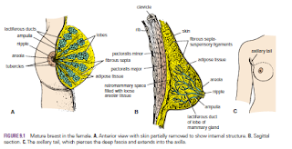
The breasts are specialized accessory glands of the skin that secrete milk. They are present in both sexes. In males and immature females, they are similar in structure. The nipples are small and surrounded by a colored area of skin called the areola. The breast tissue consists of a system of ducts embedded in connective tissue that does not extend beyond the margin of the areola.

Puberty
At puberty in females, the breasts gradually enlarge and assume their hemispherical shape under the influence of the ovarian hormones. The ducts elongate, but the increased size of the glands is mainly from the deposition of fat. The base of the breast extends from the 2nd to 6th rib and from the lateral margin of the sternum to the midaxillary line. The greater part of the gland lies in the superficial fascia. A small part, called the axillary tail, extends upward and laterally, pierces the deep fascia at the lower border of the pectoralis major muscle, and enters the axilla.
Each breast consists of 15 to 20 lobes, which radiate out from the nipple. The main duct from each lobe opens separately on the summit of the nipple and possesses a dilated ampulla just before its termination. The base of the nipple is surrounded by the areola. Tiny tubercles on the areola are produced by the underlying areolar glands.
The lobes of the gland are separated by fibrous septa that serve as suspensory ligaments. Behind the breasts is a space filled by loose connective tissue called the retromammary space.

Young Women
In young women, the breasts tend to protrude forward from a circular base.


Pregnancy
Early In the early months of pregnancy, there is a rapid increase in length and branching in the duct system. The secretory alveoli develop at the ends of the smaller ducts, and the connective tissue becomes filled with expanding and budding secretory alveoli. The vascularity of the connective tissue also increases to provide adequate nourishment for the developing gland. The nipple enlarges, and the areola becomes darker and more extensive as a result of increased deposits of melanin pigment in the epidermis. The areolar glands enlarge and become more active.
Late During the second half of pregnancy, the growth process slows. The breasts, however, continue to enlarge, mostly because of the distention of the secretory alveoli with the fluid secretion called colostrum. Postweaning Once the baby has been weaned, the breasts return to their inactive state. The remaining milk is absorbed, the secretory alveoli shrink, and most of them disappear. The interlobular connective tissue thickens. The breasts and the nipples shrink and return nearly to their original size. The pigmentation of the areola fades, but the area never lightens to its original color.

Postmenopause
After the menopause, the breast atrophies. Most of the secretory alveoli disappear, leaving behind the ducts. The amount of adipose tissue may increase or decrease. The breasts tend to shrink in size and become more pendulous. The atrophy after menopause is caused by the absence of ovarian estrogens and progesterone

Blood Supply

Arteries
The branches to the breasts include the perforating branches of the internal thoracic artery and the intercostal arteries. The axillary artery also supplies the gland via its lateral thoracic and thoracoacromial branches.

Veins
The veins correspond to the arteries.


Lymph Drainage
The lymph drainage of the mammary gland is of great clinical importance because of the frequent development of cancer in the gland and the subsequent dissemination of the malignant cells along the lymph vessels to the lymph nodes.
The lateral quadrants of the breast drain into the anterior axillary or pectoral group of nodes (situated just posterior to the lower border of the pectoralis major muscle). The medial quadrants drain by means of vessels that pierce the intercostal spaces and enter the internal thoracic group of nodes (situated within the thoracic cavity along the course of the internal thoracic artery). A few lymph vessels follow the posterior intercostal arteries and drain posteriorly into the posterior intercostal nodes (situated along the course of the posterior intercostal arteries); some vessels communicate with the lymph vessels of the opposite breast and with those of the anterior abdominal wall.


Friday, 24 June 2016

Vagina-Supports of the Vagina-Blood Supply-Vulva-Nerve Supply-Vulval Infection-The Vulva and Pregnancy-Urethral Infection-Urethral Injuries-Catheterization

Vagina
The vagina not only is the female genital canal but also serves as the excretory duct for the menstrual flow from the uterus and forms part of the birth canal. This muscular tube extends upward and backward between the vulva and the uterus (see Fig. 8.4). It measures about 3 in. (8 cm) long. The cervix of the uterus pierces its anterior wall. The vaginal orifice in a virgin possesses a thin mucosal fold, called the hymen, which is perforated at its center. The upper half of the vagina lies above the pelvic floor within the pelvis between the bladder anteriorly and the rectum posteriorly; the lower half lies within the perineum between the urethra anteriorly and the anal canal posteriorly
.
Supports of the Vagina
■■ Upper third: Levatores ani muscles and transverse cervical, pubocervical, and sacrocervical ligaments
■■ Middle third: Urogenital diaphragm
■■ Lower third: Perineal body

Blood Supply
Arteries
The vaginal artery, a branch of the internal iliac artery, and the vaginal branch of the uterine artery supply the vagina.

Vulva
The term vulva is the collective name for the female external genitalia and includes the mons pubis, labia majora and minora, the clitoris, the vestibule of the vagina, the vestibular bulb, and the greater vestibular glands.

Blood Supply
Branches of the external and internal pudendal arteries on each side.

The skin of the vulva is drained into the medial group of superficial inguinal nodes.

Lymph Drainage
Medial group of superficial inguinal nodes
.
Nerve Supply
The anterior parts of the vulva are supplied by the ilioinguinal nerves and the genital branch of the genitofemoral nerves. The posterior parts of the vulva are supplied by the branches of the perineal nerves and the posterior cutaneous nerves of the thigh.

Vulval Infection
In the region of the vulva, the presence of numerous glands and ducts opening onto the surface makes this area prone to infection. The sebaceous glands of the labia majora, the ducts of the greater vestibular glands, the vagina (with its indirect communication with the peritoneal cavity), the urethra, and the paraurethral glands can all become infected. The vagina itself has no glands and is lined with stratified squamous epithelium. Provided that the pH of its interior is kept low, it is capable of resisting infection to a remarkable degree.

The Vulva and Pregnancy
An important sign in the diagnosis of pregnancy is the appearance of a bluish discoloration of the vulva and vagina as a result of venous congestion. It appears at the 8th to 12th week and increases as the pregnancy progresses.
Urethral Infection
The short length of the female urethra predisposes to ascending infection; consequently, cystitis is more common in females than in males.
Urethral Injuries
Because of the short length of the urethra, injuries are rare. In fractures of the pelvis, the urethra may be damaged by shearing forces as it emerges from the fixed urogenital diaphragm.


Catheterization
Because the female urethra is shorter, wider, and more dilatable, catheterization is much easier than in males. Moreover, the urethra is straight, and only minor resistance is felt as the catheter passes through the urethral sphincter.


Monday, 13 June 2016

Internal Thoracic Artery in the Treatment of Coronary Artery Disease- Lymph Drainage of the Thoracic Wall

Internal Thoracic Artery in the Treatment of Coronary Artery Disease
In patients with occlusive coronary disease caused by atherosclerosis, the diseased arterial segment can be bypassed by inserting a graft. The graft most commonly used is the great saphenous vein of the leg. In some patients, the myocardium can be revascularized by surgically mobilizing one of the internal thoracic arteries and joining its distal cut end to a coronary artery.


 Lymph Drainage of the Thoracic Wall

 The lymph drainage of the skin of the anterior chest wall passes to the anterior axillary lymph nodes; that from the posterior chest wall passes to the posterior axillary nodes. The lymph drainage of the intercostal spaces passes forward to the internal thoracic nodes, situated along the internal thoracic artery, and posteriorly to the posterior intercostal nodes and the para-aortic nodes in the posterior mediastinum.