Showing posts with label Chest. Show all posts

Wednesday, 15 June 2016

Chest Pain-Somatic Chest Pain-Visceral Chest Pain-Referred Chest Pain

Chest Pain
The presenting symptom of chest pain is a common problem in clinical practice. Unfortunately, chest pain is a symptom common to many conditions and may be caused by disease in the thoracic and abdominal walls or in many different thoracic and abdominal viscera. The severity of the pain is often unrelated to the seriousness of the cause. Myocardial pain may mimic esophagitis, musculoskeletal chest wall pain, and other nonlife- threatening causes. Unless the physician is astute, a patient may be discharged with a more serious condition than the symptoms indicate. It is not good enough to have a correct diagnosis only 99% of the time with chest pain. An understanding of chest pain helps the physician in the systematic consideration of the differential diagnosis


Somatic Chest Pain
Pain arising from the chest or abdominal walls is intense and discretely localized. Somatic pain arises in sensory nerve endings in these structures and is conducted to the central nervous system by segmental spinal nerves.

Visceral Chest Pain
Visceral pain is diffuse and poorly localized. It is conducted to the central nervous system along afferent autonomic nerves. Most visceral pain fibers ascend to the spinal cord along sympathetic nerves and enter the cord through the posterior nerve roots of segmental spinal nerves. Some pain fibers from the pharynx and upper part of the esophagus and the trachea enter the central nervous system through the parasympathetic nerves via the glossopharyngeal and vagus nerves.


Referred Chest Pain
Referred chest pain is the feeling of pain at a location other than the site of origin of the stimulus, but in an area supplied by the same or adjacent segments of the spinal cord. Both somatic and visceral structures can produce referred pain.


Tuesday, 14 June 2016

Clinical Examination of the Chest-Inspection-Palpation-Percussion-Auscultation

Clinical Examination of the Chest
As medical personnel, you will be examining the chest to detect evidence of disease. Your examination consists of inspection, palpation, percussion, and auscultation


Inspection
 shows the configuration of the chest, the range of respiratory movement, and any inequalities on the two sides. The type and rate of respiration are also noted.

Palpation
enables the physician to confirm the impressions gained by inspection, especially of the respiratory movements of the chest wall. Abnormal protuberances or recession of part of the chest wall is noted. Abnormal pulsations are felt and tender areas detected.

Percussion
 is a sharp tapping of the chest wall with the fingers. This produces vibrations that extend through the tissues of the thorax. Air-containing organs such as the lungs produce a resonant note; conversely, a more solid viscus such as the heart produces a dull note. With practice, it is possible to distinguish the lungs from the heart or liver by percussion.

Auscultation
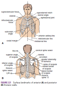
enables the physician to listen to the breath sounds as the air enters and leaves the respiratory passages. Should the alveoli or bronchi be diseased and filled with fluid, the nature of the breath sounds will be altered. The rate and rhythm of the heart can be confirmed by auscultation, and the various sounds produced by the heart and its valves during the different phases of the cardiac cycle can be heard. It may be possible to detect friction sounds produced by the rubbing together of diseased layers of pleura or pericardium. To make these examinations, the physician must be familiar with the normal structure of the thorax and must have a mental image of the normal position of the lungs and heart in relation to identifiable surface landmarks. Furthermore, it is essential that the physician be able to relate any abnormal findings to easily identifiable bony landmarks so that he or she can accurately record and communicate them to colleagues. Since the thoracic wall actively participates in the movements of respiration, many bony landmarks change their levels with each phase of respiration. In practice, to simplify matters, the levels given are those usually found at about midway between full inspiration and full expiration.


Monday, 13 June 2016

Traumatic Injury to the Back of the Chest- Traumatic Injury to the Abdominal Viscera and the Chest-Flail Chest

Traumatic Injury to the Back of the Chest
The posterior wall of the chest in the midline is formed by the vertebral column. In severe posterior chest injuries, the possibility of a vertebral fracture with associated injury to the spinal cord should be considered. Remember also the presence of the scapula, which overlies the upper seven ribs. This bone is covered with muscles and is fractured only in cases of severe trauma.

Traumatic Injury to the Abdominal Viscera and the Chest
When the anatomy of the thorax is reviewed, it is important to remember that the upper abdominal organs—namely, the liver, stomach, and spleen—may be injured by trauma to the rib cage. In fact, any injury to the chest below the level of the nipple line may involve abdominal organs as well as chest organs.

Flail Chest
In severe crush injuries, a number of ribs may break. If limited to one side, the fractures may occur near the rib angles and anteriorly near the costochondral junctions. This causes flail chest, in which a section of the chest wall is disconnected to the rest of the thoracic wall. If the fractures occur on either side of the sternum, the sternum may be flail. In either case, the stability of the chest wall is lost, and the flail segment is sucked in during inspiration and driven out during expiration, producing paradoxical and ineffective respiratory movements