Showing posts with label Traumatic. Show all posts

Sunday, 19 June 2016

Ureter- Ureteric Stones-Renal Colic-Traumatic Ureteral Injuries

Ureter
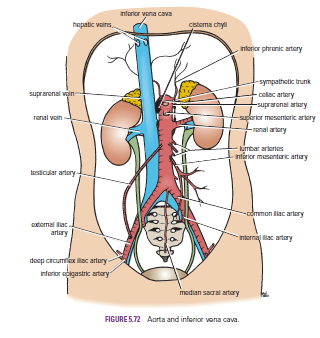
Each ureter measures about 10 in. (25 cm) long,having three constrictions along its course where the renal pelvis joins the ureter, where it is kinked as it crosses the pelvic brim, and where it pierces the bladder wall


Ureteric Stones
There are three sites of anatomic narrowing of the ureter where stones may be arrested, namely, the pelviureteral junction, the pelvic brim, and where the ureter enters the bladder. Most stones, although radiopaque, are small enough to be impossible to see definitely along the course of the ureter on plain radiographic examination. An intravenous pyelogram is usually necessary. The ureter runs down in front of the tips of the transverse processes of the lumbar vertebrae, crosses the region of the sacroiliac joint, swings out to the ischial spine, and then turns medially to the bladder
Renal Colic
The renal pelvis and the ureter send their afferent nerves into the spinal cord at segments T11 and 12 and L1 and 2. In renal colic, strong peristaltic waves of contraction pass down the ureter in an attempt to pass the stone onward. The spasm of the smooth muscle causes an agonizing colicky pain, which is referred to the skin areas that are supplied by these segments of the spinal cord, namely, the flank, loin, and groin.
When a stone enters the low part of the ureter, the pain is felt at a lower level and is often referred to the testis or the tip of the penis in the male and the labium majus in the female. Sometimes, ureteral pain is referred along the femoral branch of the genitofemoral nerve (L1 and 2) so that pain is experienced in the front of the thigh. The pain is often so severe that afferent pain impulses spread within the central nervous system, giving rise to nausea.

Traumatic Ureteral Injuries
Because of its protected position and small size, injuries to the ureter are rare. Most injuries are caused by gunshot wounds and, in a few individuals, penetrating stab wounds. Because the ureters are retroperitoneal in position, urine may escape into the retroperitoneal tissues on the posterior abdominal wall.


Monday, 13 June 2016

Traumatic Injury to the Back of the Chest- Traumatic Injury to the Abdominal Viscera and the Chest-Flail Chest

Traumatic Injury to the Back of the Chest
The posterior wall of the chest in the midline is formed by the vertebral column. In severe posterior chest injuries, the possibility of a vertebral fracture with associated injury to the spinal cord should be considered. Remember also the presence of the scapula, which overlies the upper seven ribs. This bone is covered with muscles and is fractured only in cases of severe trauma.

Traumatic Injury to the Abdominal Viscera and the Chest
When the anatomy of the thorax is reviewed, it is important to remember that the upper abdominal organs—namely, the liver, stomach, and spleen—may be injured by trauma to the rib cage. In fact, any injury to the chest below the level of the nipple line may involve abdominal organs as well as chest organs.

Flail Chest
In severe crush injuries, a number of ribs may break. If limited to one side, the fractures may occur near the rib angles and anteriorly near the costochondral junctions. This causes flail chest, in which a section of the chest wall is disconnected to the rest of the thoracic wall. If the fractures occur on either side of the sternum, the sternum may be flail. In either case, the stability of the chest wall is lost, and the flail segment is sucked in during inspiration and driven out during expiration, producing paradoxical and ineffective respiratory movements


Traumatic Injury to the Thorax-Fractured Sternum-Rib Contusion-Rib Fractures

Traumatic Injury to the Thorax
Traumatic injury to the thorax is common, especially as a result of automobile accidents

Fractured Sternum
The sternum is a resilient structure that is held in position by relatively pliable costal cartilages and bendable ribs. For these reasons, fracture of the sternum is not common; however, it does occur in high-speed motor vehicle accidents. Remember that the heart lies posterior to the sternum and may be severely contused by the sternum on impact

Rib Contusion
Bruising of a rib, secondary to trauma, is the most common rib injury. In this painful condition, a small hemorrhage occurs beneath the periosteum.

 
Rib Fractures
Fractures of the ribs are common chest injuries. In children, the ribs are highly elastic, and fractures in this age group are therefore rare. Unfortunately, the pliable chest wall in the young can be easily compressed so that the underlying lungs and heart may be injured. With increasing age, the rib cage becomes more rigid, owing to the deposit of calcium in the costal cartilages, and the ribs become brittle. The ribs then tend to break at their weakest part, their angles.
The ribs prone to fracture are those that are exposed or relatively fixed. Ribs 5 through 10 are the most commonly fractured ribs. The first four ribs are protected by the clavicle and pectoral muscles anteriorly and by the scapula and its associated muscles posteriorly. The 11th and 12th ribs float and move with the force of impact.
Because the rib is sandwiched between the skin externally and the delicate pleura internally, it is not surprising that the jagged ends of a fractured rib may penetrate the lungs and present as a pneumothorax.

Severe localized pain is usually the most important symptom of a fractured rib. The periosteum of each rib is innervated by the intercostal nerves above and below the rib. To encourage the patient to breathe adequately, it may be necessary to relieve the pain by performing an intercostal nerve block.