Showing posts with label Injury. Show all posts

Wednesday, 29 June 2016

Arterial Injury of the upper limb-Palpation and Compression of Arteries-Allen Test-Arterial Innervation and Raynaud’s Disease-

Arterial Injury of the upper limb
The arteries of the upper limb can be damaged by penetrating wounds or may require ligation in amputation operations.
Because of the existence of an adequate collateral circulation around the shoulder, elbow, and wrist joints, ligation of the main arteries of the upper limb is not followed by tissue necrosis or gangrene, provided, of course, that the arteries forming the collateral circulation are not diseased and the patient’s general circulation is satisfactory. Nevertheless, it can take days or weeks for the collateral vessels to open sufficiently to provide the distal part of the limb with the same volume of blood as previously supplied by the main artery.

Palpation and Compression of Arteries
A clinician must know where the arteries of the upper limb can be palpated or compressed in an emergency. The subclavian artery, as it crosses the first rib to become the axillary artery, can be palpated in the root of the posterior triangle of the neck. The artery can be compressed here against the first rib to stop a catastrophic hemorrhage. The third part of the axillary artery can be felt in the axilla as it lies in front of the teres major muscle. The brachial artery can be palpated in the arm as it lies on the brachialis and is overlapped from the lateral side by the biceps brachii.
The radial artery lies superficially in front of the distal end of the radius, between the tendons of the brachioradialis and flexor carpi radialis; it is here that the clinician takes the radial pulse. If the pulse cannot be felt, try feeling for the radial artery on the other wrist; occasionally, a congenitally abnormal radial artery can be difficult to feel. The radial artery can be less easily felt as it crosses the anatomic snuffbox.
The ulnar artery can be palpated as it crosses anterior to the flexor retinaculum in company with the ulnar nerve. The artery lies lateral to the pisiform bone, separated from it by the ulnar nerve. The artery is commonly damaged here in laceration wounds in front of the wrist.

Allen Test
The Allen test is used to determine the patency of the ulnar and radial arteries. With the patient’s hands resting in the lap, compress the radial arteries against the anterior surface of each radius and ask the patient to tightly clench the fists. The clenching of the fists closes off the superficial and deep palmar arterial arches. When the patient is asked to open the hands, the skin of the palms is at first white, and then normally the blood quickly flows into the arches through the ulnar arteries, causing the palms to promptly turn pink. This establishes that the ulnar arteries are patent. The patency of the radial arteries can be established by repeating the test but this time compressing the ulnar arteries as they lie lateral to the pisiform bones.

Arterial Innervation and Raynaud’s Disease
The arteries of the upper limb are innervated by sympathetic nerves. The preganglionic fibers originate from cell bodies in the 2nd to 8th thoracic segments of the spinal cord. They ascend in the sympathetic trunk and synapse in the middle cervical, inferior cervical, 1st thoracic, or stellate ganglia. The postganglionic fibers join the nerves that form the brachial plexus and are distributed to the arteries within the branches of the plexus.
For example, the digital arteries of the fingers are supplied by postganglionic sympathetic fibers that run in the digital nerves. Vasospastic diseases involving digital arterioles, such as Raynaud’s disease, may require a cervicodorsal preganglionic sympathectomy to prevent necrosis of the fingers. The operation is followed by arterial vasodilatation, with consequent increased blood flow to the upper limb.


Friday, 24 June 2016

Vaginal Examination-Injury to the Perineum during Childbirth-Pudendal Nerve Block-Area of Anesthesia-Indications-Transvaginal Procedure-Perineal Procedure

Vaginal Examination
Digital examination of the vagina may provide the physician with much valuable information concerning the health of the vaginal walls, the uterus, and the surrounding structures
Injury to the Perineum during Childbirth
The perineal body is a wedge of fibromuscular tissue that lies between the lower part of the vagina and the anal canal. It is held in position by the insertion of the perineal muscles and by the attachment of the levator ani muscles. In the female, it is a much larger structure than in the male, and it serves to support the posterior wall of the vagina. Damage by laceration during childbirth can be followed by permanent weakness of the pelvic floor.

 Few women escape some injury to the birth canal during delivery. In most, this is little more than an abrasion of the posterior vaginal wall. Spontaneous delivery of the child with the patient unattended can result in a severe tear of the lower third of the posterior wall of the vagina, the perineal body, and overlying skin. In severe tears, the lacerations may extend backward into the anal canal and damage the external sphincter. In these cases, it is imperative that an accurate repair of the walls of the anal canal, vagina, and perineal body be undertaken as soon as possible.

In the management of childbirth, when it is obvious to the obstetrician that the perineum will tear before the baby’s head emerges through the vaginal orifice, a planned surgical incision is made through the perineal skin in a posterolateral direction to avoid the anal sphincters. This procedure is known as an episiotomy. Breech deliveries and forceps deliveries are usually preceded by an episiotomy
.
Pudendal Nerve Block
Area of Anesthesia
The area anesthetized is the skin of the perineum; this nerve block does not, however, abolish sensation from the anterior part of the perineum, which is innervated by the ilioinguinal nerve and the genitofemoral nerve. Needless to say, it does not abolish pain from uterine contractions that ascend to the spinal cord via the sympathetic afferent nerves.


Indications
During the second stage of a difficult labor, when the presenting part of the fetus, usually the head, is descending through the vulva, forceps delivery and episiotomy may be necessary
.
Transvaginal Procedure
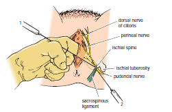
The bony landmark used is the ischial spine . The index finger is inserted through the vagina to palpate the ischial spine. The needle of the syringe is then passed through the vaginal mucous membrane toward the ischial spine. On passing through the sacrospinous ligament, the anesthetic solution is injected around the pudendal nerve .

Perineal Procedure
The bony landmark is the ischial tuberosity . The tuberosity is palpated subcutaneously through the buttock, and the needle is introduced into the pudendal canal along the medial side of the tuberosity. The canal lies about 1 in. (2.5 cm) deep to the free surface of the ischial tuberosity. The local anesthetic is then infiltrated around the pudendal nerve.


Tuesday, 21 June 2016

Palpation of the Urinary Bladder-Bladder Distention-Urinary Retention-Suprapubic Aspiration-Cystoscopy-Bladder Injuries-Difficulty with Micturition after Spinal Cord Injury-

Palpation of the Urinary Bladder
The full bladder in the adult projects up into the abdomen and may be palpated through the anterior abdominal wall above the symphysis pubis.
Bimanual palpation of the empty bladder with or without a general anesthetic is an important method of examining the bladder. In the male, one hand is placed on the anterior abdominal wall above the symphysis pubis, and the gloved index finger of the other hand is inserted into the rectum. From their knowledge of anatomy, students can see that the bladder wall can be palpated between the examining fingers. In the female, an abdominovaginal examination can be similarly made. In the child, the bladder is in a higher position than in the adult because of the relatively smaller size of the pelvis.

 
Bladder Distention
The normal adult bladder has a capacity of about 500 mL. In the presence of urinary obstruction in males, the bladder may become greatly distended without permanent damage to the bladder wall; in such cases, it is routinely possible to drain 1000 to 1200 mL of urine through a catheter.

Urinary Retention
In adult males, urinary retention is commonly caused by obstruction to the urethra by a benign or malignant enlargement of the prostate. An acute urethritis or prostatitis can also be responsible. Acute retention occurs much less frequently in females. The only anatomic cause of urinary retention in females is acute inflammation around the urethra (e.g., from herpes).

 
Suprapubic Aspiration
As the bladder fills, the superior wall rises out of the pelvis and peels the peritoneum off the posterior surface of the anterior abdominal wall. In cases of acute retention of urine, when catheterization has failed, it is possible to pass a needle into the bladder through the anterior abdominal wall above the symphysis pubis, without entering the peritoneal cavity. This is a simple method of draining off the urine in an emergency.


Cystoscopy
The mucous membrane of the bladder, the two ureteric orifices, and the urethral meatus can easily be observed by means of a cystoscope. With the bladder distended with fluid, an illuminated tube fitted with lenses is introduced into the bladder through the urethra. Over the trigone, the mucous membrane is pink and smooth. If the bladder is partially emptied, the mucous membrane over the trigone remains smooth, but it is thrown into folds elsewhere. The ureteric orifices are slitlike and eject a drop of urine at intervals of about 1 minute. The interureteric ridge and the uvula vesicae can easily be recognized.

 
Bladder Injuries
The bladder may rupture intraperitoneally or extraperitoneally. Intraperitoneal rupture usually involves the superior wall of the bladder and occurs most commonly when the bladder is full and has extended up into the abdomen. Urine and blood escape freely into the peritoneal cavity. Extraperitoneal rupture involves the anterior part of the bladder wall below the level of the peritoneal reflection; it most commonly occurs in fractures of the pelvis when bony fragments pierce the bladder wall. Lower abdominal pain and blood in the urine (hematuria) are found in most patients.
In young children, the bladder is an abdominal organ, so abdominal trauma can injure the empty bladder.

Difficulty with Micturition after Spinal Cord Injury
After injuries to the spinal cord, the nervous control of micturition is disrupted.
The normal bladder is innervated as follows:
■■ Sympathetic outflow is from the first and second lumbar segments of the spinal cord. The sympathetic nerves  inhibit contraction of the detrusor muscle of the bladder wall and stimulate closure of the sphincter vesicae.
■■ Parasympathetic outflow is from the second, third, and fourth sacral segments of the spinal cord. The parasympathetic nerves stimulate the contraction of the detrusor muscle of the bladder wall and inhibit the action of the sphincter vesicae.
■■ Sensory nerve fibers enter the spinal cord at the above segments.


Monday, 13 June 2016

Traumatic Injury to the Back of the Chest- Traumatic Injury to the Abdominal Viscera and the Chest-Flail Chest

Traumatic Injury to the Back of the Chest
The posterior wall of the chest in the midline is formed by the vertebral column. In severe posterior chest injuries, the possibility of a vertebral fracture with associated injury to the spinal cord should be considered. Remember also the presence of the scapula, which overlies the upper seven ribs. This bone is covered with muscles and is fractured only in cases of severe trauma.

Traumatic Injury to the Abdominal Viscera and the Chest
When the anatomy of the thorax is reviewed, it is important to remember that the upper abdominal organs—namely, the liver, stomach, and spleen—may be injured by trauma to the rib cage. In fact, any injury to the chest below the level of the nipple line may involve abdominal organs as well as chest organs.

Flail Chest
In severe crush injuries, a number of ribs may break. If limited to one side, the fractures may occur near the rib angles and anteriorly near the costochondral junctions. This causes flail chest, in which a section of the chest wall is disconnected to the rest of the thoracic wall. If the fractures occur on either side of the sternum, the sternum may be flail. In either case, the stability of the chest wall is lost, and the flail segment is sucked in during inspiration and driven out during expiration, producing paradoxical and ineffective respiratory movements