Showing posts with label Pain. Show all posts

Tuesday, 28 June 2016

Shoulder Joint-Movements-Stability of the Shoulder Joint-Dislocations of the Shoulder Joint-Anterior Inferior Dislocation--Posterior Dislocations-Shoulder Pain

Shoulder Joint
■■ Articulation: This occurs between the rounded head of the humerus and the shallow, pear-shaped glenoid cavity of the scapula. The articular surfaces are covered by hyaline articular cartilage, and the glenoid cavity is deepened by the presence of a fibrocartilaginous rim called the glenoid labrum.
■■ Type: Synovial ball-and-socket joint
■■ Capsule: This surrounds the joint and is attached medially to the margin of the glenoid cavity outside the labrum; laterally, it is attached to the anatomic neck of the humerus. The capsule is thin and lax, allowing a wide range of movement. It is strengthened by fibrous slips from the tendons of the subscapularis, supraspinatus, infraspinatus, and teres minor muscles (the rotator cuff muscles).
■■ Ligaments: The glenohumeral ligaments are three weak bands of fibrous tissue that strengthen the front of the capsule. The transverse humeral ligament strengthens the capsule and bridges the gap between the two tuberosities . The coracohumeral ligament strengthens the capsule above and stretches from the root of the coracoid process to the greater tuberosity of the humerus.
■■ Accessory ligaments: The coracoacromial ligament extends between the coracoid process and the acromion. Its function is to protect the superior aspect of the joint
■■ Synovial membrane: This lines the capsule and is attached to the margins of the cartilage covering the articular surfaces. It forms a tubular sheath around the tendon of the long head of the biceps brachii. It extends through the anterior wall of the capsule to form the subscapularis bursa beneath the subscapularis muscle .
■■ Nerve supply: The axillary and suprascapular nerves

Movements
The shoulder joint has a wide range of movement, and the stability of the joint has been sacrificed to permit this.
(Compare with the hip joint, which is stable but limited in its movements.) The strength of the joint depends on the tone of the short rotator cuff muscles that cross in front, above, and behind the joint—namely, the subscapularis, supraspinatus, infraspinatus, and teres minor. When the joint is abducted, the lower surface of the head of the humerus is supported by the long head of the triceps, which bows downward because of its length and gives little actual support to the humerus. In addition, the inferior part of the capsule is the weakest area.

Stability of the Shoulder Joint
The shallowness of the glenoid fossa of the scapula and the lack of support provided by weak ligaments make this joint an unstable structure. Its strength almost entirely depends on the tone of the short muscles that bind the upper end of the humerus to the scapula—namely, the subscapularis in front, the supraspinatus above, and the infraspinatus and teres minor behind. The tendons of these muscles are fused to the underlying capsule of the shoulder joint. Together, these tendons form the rotator cuff.
The least supported part of the joint lies in the inferior location, where it is unprotected by muscles.

Dislocations of the Shoulder Joint
The shoulder joint is the most commonly dislocated large joint.

Anterior Inferior Dislocation
Sudden violence applied to the humerus with the joint fully abducted tilts the humeral head downward onto the inferior weak part of the capsule, which tears, and the humeral head comes to lie inferior to the glenoid fossa. During this movement, the acromion has acted as a fulcrum. The strong flexors and adductors of the shoulder joint now usually pull the humeral head forward and upward into the subcoracoid position.

Posterior Dislocations
Posterior dislocations are rare and are usually caused by direct violence to the front of the joint. On inspection of the patient with shoulder dislocation, the rounded appearance of the shoulder is seen to be lost because the greater tuberosity of the humerus is no longer bulging laterally beneath the deltoid muscle. A subglenoid displacement of the head of the humerus into the quadrangular space can cause damage to the axillary nerve, as indicated by paralysis of the deltoid muscle and loss of skin sensation over the lower half of the deltoid. Downward displacement of the humerus can also stretch and damage the radial nerve.


Shoulder Pain

The synovial membrane, capsule, and ligaments of the shoulder joint are innervated by the axillary nerve and the suprascapular nerve. The joint is sensitive to pain, pressure, excessive traction, and distention. The muscles surrounding the joint undergo reflex spasm in response to pain originating in the joint, which in turn serves to immobilize the joint and thus reduce the pain.
Injury to the shoulder joint is followed by pain, limitation of movement, and muscle atrophy owing to disuse. It is important to appreciate that pain in the shoulder region can be caused by disease elsewhere and that the shoulder joint may be normal; for example, diseases of the spinal cord and vertebral column and the pressure of a cervical rib (see page XXX) can cause shoulder pain. Irritation of the diaphragmatic pleura or peritoneum can produce referred pain via the phrenic and supraclavicular nerves.


Sunday, 19 June 2016

Sacral Plexus-Pressure from the Fetal Head-Invasion by Malignant Tumors-Referred Pain from the Obturator Nerve-Caudal Anesthesia (Analgesia)

Sacral Plexus
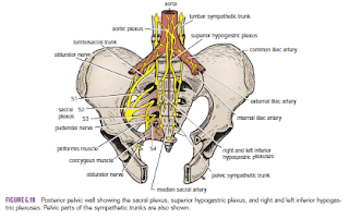
The sacral plexus lies in front of the piriformis muscle on the posterior pelvic wall. It is formed from the anterior rami of the 4th and 5th lumbar nerves and the anterior rami of the first, second, third, and fourth sacral nerves. The fourth lumbar nerve joins the fifth lumbar nerve to form the lumbosacral trunk. The lumbosacral trunk passes down into the pelvis and joins the sacral nerves as they emerge from the anterior sacral foramina.

 
Pressure from the Fetal Head
when the fetal head has descended into the pelvis During the later stages of pregnancy, , the mother often complains of discomfort or aching pain extending down one of the lower limbs. The discomfort, caused by pressure from the fetal head, is often relieved by changing position, such as lying on the side in bed.

Invasion by Malignant Tumors
The nerves of the sacral plexus can become invaded by malignant tumors extending from neighboring viscera. A carcinoma of the rectum, for example, can cause severe intractable pain down the lower limbs.

Referred Pain from the Obturator Nerve
The obturator nerve lies on the lateral wall of the pelvis and supplies the parietal peritoneum. An inflamed appendix hanging down into the pelvic cavity could cause irritation of the obturator nerve endings, leading to referred pain down the inner side of the right thigh. Inflammation of the ovaries can produce similar symptoms.

Caudal Anesthesia (Analgesia)
Anesthetic solutions can be injected into the sacral canal through the sacral hiatus. The solutions then act on the spinal roots of the 2nd, 3rd, 4th and 5th sacral and coccygeal segments of the cord as they emerge from the dura mater. The roots of higher spinal segments can also be blocked by this method. The needle must be confined to the lower part of the sacral canal, because the meninges extend down as far as the lower border of the second sacral vertebra. Caudal anesthesia is used in obstetrics to block pain fibers from the cervix of the uterus and to anesthetize the perineum


Abdominal Pain-Visceral Abdominal Pain-Somatic Abdominal Pain-Referred Abdominal Pain-Lumbar Sympathectomy-

Abdominal Pain
We will talk about Abdominal pain because it is one of the most important problems facing the physician. This section provides an anatomic basis for the different forms of abdominal pain found in clinical practice. Three distinct forms of pain exist:

 
visceral, somatic, and referred pain.
Visceral Abdominal Pain
Visceral abdominal pain means pain in abdominal organs, visceral peritoneum, and the mesenteries. The sensations that arise in viscera reach the central nervous system in afferent nerves that accompany the sympathetic nerves and enter the spinal cord through the posterior roots.
Visceral pain from the stomach is commonly referred to the epigastrium The causes of visceral pain include impaired blood supply (ischemia) to a viscus, stretching of a viscus or mesentery, distention of a hollow viscus, and chemical damage  to a viscus or its covering peritoneum. Pain arising from an abdominal viscus is dull and poorly localized. Visceral pain is referred to the midline, probably because the viscera develop embryologically as midline structures and receive a bilateral nerve supply; many viscera later move laterally as development proceeds, taking their nerve supply with them.

violent contraction of smooth muscle cause Colic  is a form of visceral pain; it is commonly caused by luminal obstruction as in intestinal obstruction, in the passage of a gallstone in the biliary ducts, or in the passage of a stone in the ureters.

Many visceral afferent fibers that enter the spinal cord participate in reflex activity. Reflex sweating, salivation, nausea, vomiting, and increased heart rate may accompany visceral pain.


Somatic Abdominal Pain
Somatic abdominal pain in the abdominal wall can arise from the skin, fascia, muscles, and parietal peritoneum. It can be severe and precisely localized. When the origin is on one side of the midline, the pain is also lateralized. The somatic pain impulses from the abdomen reach the central nervous system in the following segmental spinal nerves:
■■ Central part of the diaphragm: Phrenic nerve (C3, 4, and 5)
■■ Peripheral part of the diaphragm: Intercostal nerves(T7 to 11)
■■ Anterior abdominal wall: Thoracic nerves (T7 to 12) and the1st lumbar nerve
■■ Pelvic wall: Obturator nerve (L2, 3, and 4)

The inflamed parietal peritoneum is extremely sensitive, and because the full thickness of the abdominal wall is innervated by the same nerves, it is not surprising to find cutaneous hypersensitivity (hyperesthesia) and tenderness. Local reflexes involving the same nerves bring about a protective phenomenon in which the abdominal muscles increase in tone. This increased tone or rigidity, sometimes called guarding, is an attempt to rest and localize the inflammatory process. Rebound tenderness occurs when the parietal peritoneum is inflamed. Any movement of that inflamed peritoneum, even when that movement is elicited by removing the examining hand from a site distant from the inflamed peritoneum, brings about tenderness.
Examples of acute, severe, localized pain originating in the parietal peritoneum are seen in the later stages of appendicitis. Cutaneous hyperesthesia, tenderness, and muscular spasm or rigidity occur in the lower right quadrant of the anterior abdominal wall. A perforated peptic ulcer, in which the parietal peritoneum is chemically irritated, produces the same symptoms and signs but involves the right upper and lower quadrants.


Referred Abdominal Pain
Referred abdominal pain is the feeling of pain at a location other than the site of origin of the stimulus but in an area supplied by the same or adjacent segments of the spinal cord. Both somatic and visceral structures can produce referred pain. In the case of referred somatic pain, the possible explanation is that the nerve fibers from the diseased structure and the area where the pain is felt ascend in the central nervous system along a common pathway, and the cerebral cortex is incapable of distinguishing between the sites. Examples of referred somatic pain follow. Pleurisy involving the lower part of the costal parietal pleura can give rise to referred pain in the abdomen because the lower parietal pleura receives its sensory innervation from the lower five intercostal nerves, which also innervate the skin and muscles of the anterior abdominal wall.

 
Lumbar Sympathectomy
Lumbar sympathectomy is performed mainly to produce a vasodilatation of the arteries of the lower limb in patients with vasospastic disorders. The preganglionic sympathetic fibers that supply the vessels of the lower limb leave the spinal cord from segments T11 to L2. They synapse in the lumbar and sacral ganglia of the sympathetic trunks. The postganglionic fibers join the lumbar and sacral nerves and are distributed to the vessels of the limb as branches of these nerves. Additional postganglionic fibers pass directly from the lumbar ganglia to the common and external iliac arteries, but they follow the latter artery only down as far as the inguinal ligament. In the male, a bilateral lumbar sympathectomy may be followed by loss of ejaculatory power, but erection is not impaired.


Kidneys-Renal Mobility-Kidney Trauma-Kidney Tumors-Renal Pain-Transplanted Kidneys-

Kidneys
The kidneys function is to excrete most of the waste products of metabolism. alsoThey play a major role in controlling the water and electrolyte balance within the body and in maintaining the acid–base balance of the blood. The waste products leave the kidneys as urine, which passes down the ureters to the urinary bladder, located within the pelvis. The urine leaves the body in the urethra.
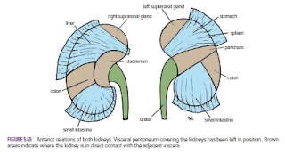
The kidneys are reddish brown and lie behind the peritoneum high up on the posterior abdominal wall on either side of the vertebral column; they are largely under cover of the costal margin.
The right kidney lies slightly lower than the left kidney because of the large size of the right lobe of the liver. With contraction of the diaphragm during respiration, both kidneys move downward in a vertical direction by as much as 1 in. (2.5 cm). On the medial concave border of each kidney is a vertical slit that is bounded by thick lips of renal substance and is called the hilum. The hilum extends into a large cavity called the renal sinus. The hilum transmits, from the front backward, the renal vein, two branches of the renal artery, the ureter, and the third branch of the renal artery (VAUA). Lymph vessels and sympathetic fibers also pass through the hilum

Renal Mobility
The kidneys are maintained in their normal position by intraabdominal pressure and by their connections with the perirenal fat and renal fascia. Each kidney moves slightly with respiration. The right kidney lies at a slightly lower level than the left kidney, and the lower pole may be palpated in the right lumbar region at the end of deep inspiration in a person with poorly developed abdominal musculature. Should the amount of perirenal fat be reduced, the mobility of the kidney may become excessive and produce symptoms of renal colic caused by kinking of the ureter. Excessive mobility of the kidney leaves the suprarenal gland undisturbed because the latter occupies a separate compartment in the renal fascia.


Kidney Trauma
The kidneys are well protected by the lower ribs, the lumbar muscles, and the vertebral column. However, a severe blunt injury applied to the abdomen may crush the kidney against the last rib and the vertebral column. Depending on the severity of the blow, the injury varies from a mild bruising to a complete laceration of the organ. Penetrating injuries are usually caused by stab wounds or gunshot wounds and often involve other viscera. Because 25% of the cardiac outflow passes through the kidneys, renal injury can result in rapid blood loss


Kidney Tumors
Malignant tumors of the kidney have a strong tendency to spread along the renal vein. The left renal vein receives the left testicular vein in the male, and this may rarely become blocked, producing left-sided varicocele.

Renal Pain
Renal pain varies from a dull ache to a severe pain in the flank that may radiate downward into the lower abdomen. Renal pain can result from stretching of the kidney capsule or spasm of the smooth muscle in the renal pelvis. The afferent nerve fibers pass through the renal plexus around the renal artery and ascend to the spinal cord through the lowest splanchnic nerve in the thorax and the sympathetic trunk. They enter the spinal cord at the level of T12. Pain is commonly referred along the distribution of the subcostal nerve (T12) to the flank and the anterior abdominal wall.

Transplanted Kidneys
The iliac fossa on the posterior abdominal wall is the usual site chosen for transplantation of the kidney. The fossa is exposed through an incision in the anterior abdominal wall just above the inguinal ligament. The iliac fossa in front of the iliacus muscle is approached retroperitoneally. The kidney is positioned and the vascular anastomosis constructed. The renal artery is anastomosed end to end to the internal iliac artery and the renal vein is anastomosed end to side to the external iliac vein. Anastomosis of the branches of the internal iliac arteries on the two sides is sufficient so that the pelvic viscera on the side of the renal arterial anastomosis are not at risk. Ureterocystostomy is then performed by opening the bladder and providing a wide entrance of the ureter through the bladder wall.