Showing posts with label Pregnancy. Show all posts

Sunday, 26 June 2016

The Breasts-The Breasts at Puberty-Young Women-The Breast atPregnancy-Postmenopause-Blood Supply to Breast-Arteries-Veins-Lymph Drainage-

The Breasts
The breasts, they are situated in the pectoral region so they are not anatomically part of the upper limb and their blood supply and lymphatic drainage is largely into the armpit. Their clinical importance cannot be overemphasized.
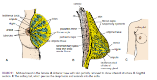
The breasts are specialized accessory glands of the skin that secrete milk. They are present in both sexes. In males and immature females, they are similar in structure. The nipples are small and surrounded by a colored area of skin called the areola. The breast tissue consists of a system of ducts embedded in connective tissue that does not extend beyond the margin of the areola.

Puberty
At puberty in females, the breasts gradually enlarge and assume their hemispherical shape under the influence of the ovarian hormones. The ducts elongate, but the increased size of the glands is mainly from the deposition of fat. The base of the breast extends from the 2nd to 6th rib and from the lateral margin of the sternum to the midaxillary line. The greater part of the gland lies in the superficial fascia. A small part, called the axillary tail, extends upward and laterally, pierces the deep fascia at the lower border of the pectoralis major muscle, and enters the axilla.
Each breast consists of 15 to 20 lobes, which radiate out from the nipple. The main duct from each lobe opens separately on the summit of the nipple and possesses a dilated ampulla just before its termination. The base of the nipple is surrounded by the areola. Tiny tubercles on the areola are produced by the underlying areolar glands.
The lobes of the gland are separated by fibrous septa that serve as suspensory ligaments. Behind the breasts is a space filled by loose connective tissue called the retromammary space.

Young Women
In young women, the breasts tend to protrude forward from a circular base.


Pregnancy
Early In the early months of pregnancy, there is a rapid increase in length and branching in the duct system. The secretory alveoli develop at the ends of the smaller ducts, and the connective tissue becomes filled with expanding and budding secretory alveoli. The vascularity of the connective tissue also increases to provide adequate nourishment for the developing gland. The nipple enlarges, and the areola becomes darker and more extensive as a result of increased deposits of melanin pigment in the epidermis. The areolar glands enlarge and become more active.
Late During the second half of pregnancy, the growth process slows. The breasts, however, continue to enlarge, mostly because of the distention of the secretory alveoli with the fluid secretion called colostrum. Postweaning Once the baby has been weaned, the breasts return to their inactive state. The remaining milk is absorbed, the secretory alveoli shrink, and most of them disappear. The interlobular connective tissue thickens. The breasts and the nipples shrink and return nearly to their original size. The pigmentation of the areola fades, but the area never lightens to its original color.

Postmenopause
After the menopause, the breast atrophies. Most of the secretory alveoli disappear, leaving behind the ducts. The amount of adipose tissue may increase or decrease. The breasts tend to shrink in size and become more pendulous. The atrophy after menopause is caused by the absence of ovarian estrogens and progesterone

Blood Supply

Arteries
The branches to the breasts include the perforating branches of the internal thoracic artery and the intercostal arteries. The axillary artery also supplies the gland via its lateral thoracic and thoracoacromial branches.

Veins
The veins correspond to the arteries.


Lymph Drainage
The lymph drainage of the mammary gland is of great clinical importance because of the frequent development of cancer in the gland and the subsequent dissemination of the malignant cells along the lymph vessels to the lymph nodes.
The lateral quadrants of the breast drain into the anterior axillary or pectoral group of nodes (situated just posterior to the lower border of the pectoralis major muscle). The medial quadrants drain by means of vessels that pierce the intercostal spaces and enter the internal thoracic group of nodes (situated within the thoracic cavity along the course of the internal thoracic artery). A few lymph vessels follow the posterior intercostal arteries and drain posteriorly into the posterior intercostal nodes (situated along the course of the posterior intercostal arteries); some vessels communicate with the lymph vessels of the opposite breast and with those of the anterior abdominal wall.


Friday, 24 June 2016

Vagina-Supports of the Vagina-Blood Supply-Vulva-Nerve Supply-Vulval Infection-The Vulva and Pregnancy-Urethral Infection-Urethral Injuries-Catheterization

Vagina
The vagina not only is the female genital canal but also serves as the excretory duct for the menstrual flow from the uterus and forms part of the birth canal. This muscular tube extends upward and backward between the vulva and the uterus (see Fig. 8.4). It measures about 3 in. (8 cm) long. The cervix of the uterus pierces its anterior wall. The vaginal orifice in a virgin possesses a thin mucosal fold, called the hymen, which is perforated at its center. The upper half of the vagina lies above the pelvic floor within the pelvis between the bladder anteriorly and the rectum posteriorly; the lower half lies within the perineum between the urethra anteriorly and the anal canal posteriorly
.
Supports of the Vagina
■■ Upper third: Levatores ani muscles and transverse cervical, pubocervical, and sacrocervical ligaments
■■ Middle third: Urogenital diaphragm
■■ Lower third: Perineal body

Blood Supply
Arteries
The vaginal artery, a branch of the internal iliac artery, and the vaginal branch of the uterine artery supply the vagina.

Vulva
The term vulva is the collective name for the female external genitalia and includes the mons pubis, labia majora and minora, the clitoris, the vestibule of the vagina, the vestibular bulb, and the greater vestibular glands.

Blood Supply
Branches of the external and internal pudendal arteries on each side.

The skin of the vulva is drained into the medial group of superficial inguinal nodes.

Lymph Drainage
Medial group of superficial inguinal nodes
.
Nerve Supply
The anterior parts of the vulva are supplied by the ilioinguinal nerves and the genital branch of the genitofemoral nerves. The posterior parts of the vulva are supplied by the branches of the perineal nerves and the posterior cutaneous nerves of the thigh.

Vulval Infection
In the region of the vulva, the presence of numerous glands and ducts opening onto the surface makes this area prone to infection. The sebaceous glands of the labia majora, the ducts of the greater vestibular glands, the vagina (with its indirect communication with the peritoneal cavity), the urethra, and the paraurethral glands can all become infected. The vagina itself has no glands and is lined with stratified squamous epithelium. Provided that the pH of its interior is kept low, it is capable of resisting infection to a remarkable degree.

The Vulva and Pregnancy
An important sign in the diagnosis of pregnancy is the appearance of a bluish discoloration of the vulva and vagina as a result of venous congestion. It appears at the 8th to 12th week and increases as the pregnancy progresses.
Urethral Infection
The short length of the female urethra predisposes to ascending infection; consequently, cystitis is more common in females than in males.
Urethral Injuries
Because of the short length of the urethra, injuries are rare. In fractures of the pelvis, the urethra may be damaged by shearing forces as it emerges from the fixed urogenital diaphragm.


Catheterization
Because the female urethra is shorter, wider, and more dilatable, catheterization is much easier than in males. Moreover, the urethra is straight, and only minor resistance is felt as the catheter passes through the urethral sphincter.


Uterus in the Child-Uterus after Menopause-Uterus in Pregnancy-Role of the Uterus in Labor-Prolapse of the Uterus-Hysterectomy and Damage to the Ureter-Varicosed Veins and Hemorrhoids in Pregnancy-

Uterus in the Child
The fundus and body of the uterus remain small until puberty, when they enlarge greatly in response to the estrogens secreted by the ovaries.

Uterus after Menopause
After menopause because the ovaries no longer produce estrogens and progesterone, the uterus atrophies and becomes smaller and less vascular. These changes occur.

Uterus in Pregnancy
During pregnancy, the uterus becomes greatly enlarged as a result of the increasing production of estrogens and progesterone, first by the corpus luteum of the ovary and later by the placenta. At first, it remains as a pelvic organ, but by the third month the fundus rises out of the pelvis, and by the ninth month it has reached the xiphoid process. The increase in size is largely a result of hypertrophy of the smooth muscle fibers of the myometrium, although some hyperplasia takes place.

 
Role of the Uterus in Labor
Labor, or parturition, is the series of processes by which the baby, the fetal membranes, and the placenta are expelled from the genital tract of the mother. Normally, this process takes place at the end of the 10th lunar month, at which time the pregnancy is said to be at term. The cause of the onset of labor is not definitely known. By the end of pregnancy, the contractility of the uterus has been fully developed in response to estrogen, and it is particularly sensitive to the actions of oxytocin at this time. It is possible that the onset of labor is triggered by the sudden withdrawal of progesterone. Once the presenting part (usually the fetal head) starts to stretch the cervix, it is thought that a nervous reflex mechanism is initiated and increases the force of the contractions of the uterine body. The uterine muscular activity is largely independent of the extrinsic innervation. In women in labor, spinal anesthesia does not interfere with the normal uterine contractions. Severe emotional disturbance, however, can cause premature parturition


Prolapse of the Uterus
The great importance of the tone of the levatores ani muscles in supporting the uterus. The importance of the transverse cervical, pubocervical, and sacrocervical ligaments in positioning the cervix within the pelvic cavity. Damage to these structures during childbirth or general poor body muscular tone may result in downward displacement of the uterus called uterine prolapse. It most commonly reveals itself after menopause, when the visceral pelvic fascia tends to atrophy along with the pelvic organs. In advanced cases, the cervix descends the length of the vagina and may protrude through the orifice.
Because of the attachment of the cervix to the vaginal vault, it follows that prolapse of the uterus is always accompanied by some prolapse of the vagina.

Hysterectomy and Damage to the Ureter
During the surgical procedure of hysterectomy, great care must be exercised to not damage the ureters. When the surgeon is looking for the uterine artery on each side at the base of the broad ligament, it is essential that he or she first identifies the ureter before clamping and tying off the artery. The uterine artery passes forward from the internal iliac artery and crosses the ureter at right angles to reach the cervix at the level of the internal os.


Varicosed Veins and Hemorrhoids in Pregnancy
Varicosed veins and hemorrhoids are common conditions in pregnancy. The following factors probably contribute to their cause: pressure of the gravid uterus on the inferior vena cava and the inferior mesenteric vein, impairing venous return, and increased progesterone levels in the blood, leading to relaxation of the smooth muscle in the walls of the veins and venous dilatation.


Tuesday, 21 June 2016

Uterine Tube-Uterine TubeFunction-Blood Supply-The Uterine Tube as a Conduit for Infection-Pelvic Inflammatory Disease-Ectopic Pregnancy- Tubal Ligation-

Uterine Tube
The two uterine tubes are each about 4 in. (10 cm) long and lie in the upper border of the broad ligament . Each connects the peritoneal cavity in the region of the ovary with the cavity of the uterus. The uterine tube is divided into four parts:
1. The infundibulum is the funnel-shaped lateral end that projects beyond the broad ligament and overlies the ovary. The free edge of the funnel has several fingerlike processes, known as fimbriae, which are draped over the ovary .
2. The ampulla is the widest part of the tube.
3. The isthmus is the narrowest part of the tube and lies just lateral to the uterus .
4. The intramural part is the segment that pierces the uterine wall .

Function
The uterine tube receives the ovum from the ovary and provides a site where fertilization of the ovum can take place (usually in the ampulla). It provides nourishment for the fertilized ovum and transports it to the cavity of the uterus. The tube serves as a conduit along which the spermatozoa travel to reach the ovum.

Blood Supply

Arteries
The uterine artery from the internal iliac artery and the ovarian artery from the abdominal aorta .

Veins
The veins correspond to the arteries.

Lymph Drainage
The internal iliac and para-aortic nodes.


Nerve Supply
Sympathetic and parasympathetic nerves from the inferior hypogastric plexuses

The Uterine Tube as a Conduit for Infection
The uterine tube lies in the upper free border of the broad ligament and is a direct route of communication from the vulva through the vagina and uterine cavity to the peritoneal cavity.

Pelvic Inflammatory Disease
The pathogenic organism(s) enter the body through sexual contact and ascend through the uterus and enter the uterine tubes. Salpingitis may follow, with leakage of pus into the peritoneal cavity, causing pelvic peritonitis. A pelvic abscess usually follows, or the infection spreads farther, causing general peritonitis.

Ectopic Pregnancy
Implantation and growth of a fertilized ovum may occur outside the uterine cavity in the wall of the uterine tube. This is a variety of ectopic pregnancy. There being no decidua formation in the tube, the eroding action of the trophoblast quickly destroys the wall of the tube. Tubal abortion or rupture of the tube, with the effusion of a large quantity of blood into the peritoneal cavity, is the common result.
The blood pours down into the rectouterine pouch (pouch of Douglas) or into the uterovesical pouch. The blood may quickly ascend into the general peritoneal cavity, giving rise to severe abdominal pain, tenderness, and guarding. Irritation of the subdiaphragmatic peritoneum (supplied by phrenic nerves C3, 4, and 5) may give rise to referred pain to the shoulder skin (supraclavicular nerves C3 and 4).


Tubal Ligation
Ligation and division of the uterine tubes is a method of obtaining permanent birth control and is usually restricted to women who already have children. The ova that are discharged from the ovarian follicles degenerate in the tube proximal to the obstruction. If, later, the woman wishes to have an additional child, restoration of the continuity of the uterine tubes can be attempted, and, in about 20% of women, fertilization occurs.