Showing posts with label Muscles. Show all posts

Wednesday, 6 July 2016

Ligaments of the Gluteal Region-Sacrotuberous Ligament-Sacrospinous Ligament-Greater Sciatic Foramen-Foramina of the Gluteal Region-Lesser Sciatic Foramen-Muscles of the Gluteal Region-

Ligaments of the Gluteal Region
The two important ligaments in the gluteal region are the sacrotuberous and sacrospinous ligaments. The function of these ligaments is to stabilize the sacrum and prevent its rotation at the sacroiliac joint by the weight of the vertebral column.
Sacrotuberous Ligament
The sacrotuberous ligament connects the back of the sacrum to the ischial tuberosity.
Sacrospinous Ligament
The sacrospinous ligament connects the back of the sacrum to the spine of the ischium.

Foramina of the Gluteal Region
The two important foramina in the gluteal region are the greater sciatic foramen and the lesser sciatic foramen.
Greater Sciatic Foramen
The greater sciatic foramen is formed by the greater sciatic notch of the hip bone and the sacrotuberous and sacrospinous ligaments. It provides an exit from the pelvis into the gluteal region.
The following structures exit the foramen:
■■ Piriformis
■■ Sciatic nerve
■■ Posterior cutaneous nerve of the thigh

■■ Superior and inferior gluteal nerves
■■ Nerves to the obturator internus and quadratus femoris
■■ Pudendal nerve
■■ Superior and inferior gluteal arteries and veins
■■ Internal pudendal artery and vein


Lesser Sciatic Foramen
The lesser sciatic foramen is formed by the lesser sciatic notch of the hip bone and the sacrotuberous and sacrospinous ligaments. It provides an entrance into the perineum from the gluteal region. Its presence enables nerves and blood vessels that have left the pelvis through the greater sciatic foramen above the pelvic floor to enter the perineum below the pelvic floor.
The following structures pass through the foramen
■■ Tendon of obturator internus muscle
■■ Nerve to obturator internus
■■ Pudendal nerve
■■ Internal pudendal artery and vein

Muscles of the Gluteal Region
The muscles of the gluteal region include the gluteus maximus, the gluteus medius, the gluteus minimus, the tensor fasciae latae, the piriformis, the obturator internus, the superior and inferior gemelli, and the quadratus femoris.
Note the following:
■■ The gluteus maximus is the largest muscle in the body. It lies superficial in the gluteal region and is largely responsible for the prominence of the buttock.
■■ The tensor fasciae latae runs downward and backward to its insertion in the iliotibial tract and thus assists the gluteus maximus muscle in maintaining the knee in the extended position.


Tuesday, 28 June 2016

Fascial Compartments of the Upper Arm-Contents of the Anterior Fascial Compartment of the Upper Arm-Muscles of the Anterior Fascial Compartment-Lymphangitis-Lymphadenitis-Biceps Brachii and Osteoarthritis of the Shoulder Joint-

Fascial Compartments of the Upper Arm
The upper arm is enclosed in a sheath of deep fascia. Two fascial septa, one on the medial side and one on the lateral side, extend from this sheath and are attached to the medial and lateral supracondylar ridges of the humerus, respectively. By this means, the upper arm is divided into an anterior and a posterior fascial compartment, each having its muscles, nerves, and arteries.

Contents of the Anterior Fascial Compartment of the Upper Arm
■■ Muscles: Biceps brachii, coracobrachialis, and brachialis
■■ Blood supply: Brachial artery
■■ Nerve supply to the muscles: Musculocutaneous nerve
■■ Structures passing through the compartment: Musculocutaneous, median, and ulnar nerves; brachial artery and basilic vein. The radial nerve is present in the lower part of the compartment.

Muscles of the Anterior Fascial Compartment
The muscles of the anterior fascial compartment ,Note that the biceps brachii is a powerful supinator, and this action is made use of in twisting the corkscrew into the cork or driving the screw into wood with a screwdriver. The biceps also is a powerful flexor of the elbow joint and a weak flexor of the shoulder joint.
 
Lymphangitis
Infection of the lymph vessels (lymphangitis) of the arm is common. Red streaks along the course of the lymph vessels are characteristic of the condition. The lymph vessels from the thumb and index finger and the lateral part of the hand follow the cephalic vein to the infraclavicular group of axillary nodes; those from the middle, ring, and little fingers and from the medial part of the hand follow the basilic vein to the supratrochlear node, which lies in the superficial fascia just above the medial epicondyle of the humerus, and thence to the lateral group of axillary nodes.


Lymphadenitis
Once the infection reaches the lymph nodes, they become enlarged and tender, a condition known as lymphadenitis.
Most of the lymph vessels from the fingers and palm pass to the dorsum of the hand before passing up into the forearm. This explains the frequency of inflammatory edema, or even abscess formation, which may occur on the dorsum of the hand after infection of the fingers or palm.

Biceps Brachii and Osteoarthritis of the Shoulder Joint
The tendon of the long head of biceps is attached to the supraglenoid tubercle within the shoulder joint. Advanced osteoarthritic changes in the joint can lead to erosion and fraying of the tendon by osteophytic outgrowths, and rupture of the tendon can occur.


Sternoclavicular Joint-Movements-Muscles Producing Movement-Important Relations-Sternoclavicular Joint Injuries-Anterior dislocation-Posterior dislocation-

Sternoclavicular Joint
■■ Articulation: This occurs between the sternal end of the clavicle, the manubrium sterni, and the 1st costal cartilage
■■ it s type of joints is: Synovial double-plane joint
■■ Capsule: This surrounds the joint and is attached to the margins of the articular surfaces.
■■ Ligaments: The capsule is reinforced in front of and behind the joint by the strong sternoclavicular ligaments.
■■ Articular disc: This flat fibrocartilaginous disc lies within the joint and divides the joint’s interior into two compartments. Its circumference is attached to the interior of the capsule, but it is also strongly attached to the superior margin of the articular surface of the clavicle above and to the first costal cartilage below.
■■ Accessory ligament: The costoclavicular ligament is a strong ligament that runs from the junction of the 1st rib with the 1st costal cartilage to the inferior surface of the sternal end of the clavicle.
■■ Synovial membrane: This lines the capsule and is attached to the margins of the cartilage covering the articular surfaces.
■■ Nerve supply: The supraclavicular nerve and the nerve to the subclavius muscle.

Movements
Forward and backward movement of the clavicle takes place in the medial compartment. Elevation and depression of the clavicle take place in the lateral compartment.

Muscles Producing Movement
The forward movement of the clavicle is produced by the serratus anterior muscle. The backward movement is produced by the trapezius and rhomboid muscles. Elevation of the clavicle is produced by the trapezius, sternocleidomastoid, levator scapulae, and rhomboid muscles. Depression of the clavicle is produced by the pectoralis minor and the subclavius muscles.


Important Relations
■■ Anteriorly: The skin and some fibers of the sternocleidomastoid and pectoralis major muscles
■■ Posteriorly: The sternohyoid muscle; on the right, the brachiocephalic artery; on the left, the left brachiocephalic vein and the left common carotid artery

Sternoclavicular Joint Injuries
The strong costoclavicular ligament firmly holds the medial end of the clavicle to the 1st costal cartilage. Violent forces directed along the long axis of the clavicle usually result in fracture of that bone, but dislocation of the sternoclavicular joint takes place occasionally.

Anterior dislocation
 results in the medial end of the clavicle projecting forward beneath the skin; it may also be pulled upward by the sternocleidomastoid muscle.


Posterior dislocation
 usually follows direct trauma applied to the front of the joint that drives the clavicle backward. This type is the more serious one because the displaced clavicle may press on the trachea, the esophagus, and major blood vessels in the root of the neck.
If the costoclavicular ligament ruptures completely, it is difficult to maintain the normal position of the clavicle once reduction has been accomplished.


Sunday, 26 June 2016

The Axilla-Walls of the Axilla-Contents of the Axilla-Key Muscles in the Axilla-Pectoralis Minor-Clavipectoral Fascia-Absent Pectoralis Major-

The Axilla
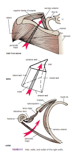
The axilla, or armpit, is a pyramid-shaped space between the upper part of the arm and the side of the chest. It forms an important passage for nerves, blood, and lymph vessels as they travel from the root of the neck to the upper limb. The upper end of the axilla, or apex, is directed into the root of the neck and is bounded in front by the clavicle, behind by the upper border of the scapula, and medially by the outer border of the first rib. The lower end, or base, is bounded in front by the anterior axillary fold (formed by the lower border of the pectoralis major muscle), behind by the posterior axillary fold (formed by the tendon of latissimus dorsi and the teres major muscle), and medially by the chest wall

Walls of the Axilla
The walls of the axilla are made up as follows:
■■ Anterior wall: By the pectoralis major, subclavius, and pectoralis minor muscles
■■ Posterior wall: By the subscapularis, latissimus dorsi, and teres major muscles from above down
■■ Medial wall: By the upper four or five ribs and the intercostal spaces covered by the serratus anterior muscle
■■ Lateral wall: By the coracobrachialis and biceps muscles in the bicipital groove of the humerus
The base is formed by the skin stretching between the anterior and posterior walls.

Contents of the Axilla
The axilla contains the axillary artery and its branches, which supply blood to the upper limb; the axillary vein and its tributaries, which drain blood from the upper limb; and lymph vessels and lymph nodes, which drain lymph from the upper limb and the breast and from the skin of the trunk, down as far as the level of the umbilicus. Lying among these structures in the axilla is an important nerve plexus, the brachial plexus, which innervates the upper limb. These structures are embedded in fat.


Key Muscles in the Axilla
Pectoralis Minor
The pectoralis minor is a thin triangular muscle that lies beneath the pectoralis major. It arises from the3rd, 4th, and 5th ribs and runs upward and laterally to be inserted by its apex into the coracoid process of the scapula. It crosses the axillary artery and the brachial plexus of nerves. It is used when describing the axillary artery to divide it into three parts
.
Clavipectoral Fascia
The clavipectoral fascia is a strong sheet of connective tissue that is attached above to the clavicle. Below, it splits to enclose the pectoralis minor muscle and then continues downward as the suspensory ligament of the axilla and joins the fascial floor of the armpit.

Absent Pectoralis Major
Occasionally, parts of the pectoralis major muscle may be absent. The sternocostal origin is the most commonly missing part, and this causes weakness in adduction and medial rotation of the shoulder joint.