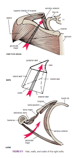
The
Axilla
The axilla, or armpit, is a pyramid-shaped space between the
upper part of the arm and the side of the chest. It forms an important passage
for nerves, blood, and lymph vessels as they travel from the root of the neck
to the upper limb. The upper end of the axilla, or apex, is directed into the
root of the neck and is bounded in front by the clavicle, behind by the upper
border of the scapula, and medially by the outer border of the first rib. The
lower end, or base, is bounded in front by the anterior axillary fold (formed
by the lower border of the pectoralis major muscle), behind by the posterior
axillary fold (formed by the tendon of latissimus dorsi and the teres major
muscle), and medially by the chest wall
Walls
of the Axilla
The walls of the axilla are made up as follows:
■■
Anterior wall: By the pectoralis major, subclavius, and pectoralis minor
muscles
■■ Posterior wall: By the subscapularis, latissimus dorsi, and teres major muscles from above down
■■ Posterior wall: By the subscapularis, latissimus dorsi, and teres major muscles from above down
■■
Medial wall: By the upper four or five ribs and the intercostal spaces covered
by the serratus anterior muscle
■■
Lateral wall: By the coracobrachialis and biceps muscles in the bicipital
groove of the humerus
The base is formed by the skin stretching between the anterior and posterior walls.
The base is formed by the skin stretching between the anterior and posterior walls.
Contents
of the Axilla
The axilla contains the axillary artery and its branches, which
supply blood to the upper limb; the axillary vein and its tributaries, which
drain blood from the upper limb; and lymph vessels and lymph nodes, which drain
lymph from the upper limb and the breast and from the skin of the trunk, down
as far as the level of the umbilicus. Lying among these structures in the
axilla is an important nerve plexus, the brachial plexus, which innervates the
upper limb. These structures are embedded in fat.
Key
Muscles in the Axilla
Pectoralis Minor
The pectoralis minor is a thin triangular muscle that lies beneath
the pectoralis major. It arises from the3rd, 4th, and 5th ribs and runs upward
and laterally to be inserted by its apex into the coracoid process of the
scapula. It crosses the axillary artery and the brachial plexus of nerves. It
is used when describing the axillary artery to divide it into three parts
.
.
Clavipectoral Fascia
The clavipectoral fascia is a strong sheet of connective tissue
that is attached above to the clavicle. Below, it splits to enclose the
pectoralis minor muscle and then continues downward as the suspensory ligament
of the axilla and joins the fascial floor of the armpit.
Absent
Pectoralis Major
Occasionally, parts of the pectoralis major muscle may be absent.
The sternocostal origin is the most commonly missing part, and this causes
weakness in adduction and medial rotation of the shoulder joint.


