Showing posts with label Cancer. Show all posts

Sunday, 19 June 2016

Partial and Complete Prolapse of the Rectum-Cancer of the Rectum-Rectal Injuries-Pelvic Appendix

Partial and Complete Prolapse of the Rectum
Partial and complete prolapses of the rectum through the anus are relatively common clinical conditions. In partial prolapse, the rectal mucous membrane and submucous coat protrude for a short distance outside the anus. In complete prolapse, the whole thickness of the rectal wall protrudes through the anus. In both conditions, many causative factors may be involved. However, damage to the levatores ani muscles as the result of childbirth and poor muscle tone in the aged are important contributing factors. A complete rectal prolapse may be regarded as a sliding hernia through the pelvic diaphragm.

 
Cancer of the Rectum
Cancer of the rectum is a common clinical finding that remains localized to the rectal wall for a considerable time. At first, it tends to spread locally in the lymphatics around the circumference of the bowel. Later, it spreads upward and laterally along the lymph vessels, following the superior rectal and middle rectal arteries. Venous spread occurs late, and because the superior rectal vein is a tributary of the portal vein, the liver is a common site for secondary deposits.

Once the malignant tumor has extended beyond the confines of the rectal wall, knowledge of the anatomic relations of the rectum will enable a physician to assess the structures and organs likely to be involved. In both sexes, a posterior penetration involves the sacral plexus and can cause severe intractable pain down the leg in the distribution of the sciatic nerve. A lateral penetration may involve the ureter. An anterior penetration in the male may involve the prostate, seminal vesicles, or bladder; in the female, the vagina and uterus may be invaded. It is clear from the anatomic features of the rectum and its lymph drainage that a wide resection of the rectum with its lymphatic field offers the best chance of cure. When the tumor has spread to contiguous organs and is of a low grade of malignancy, some form of pelvic evisceration may be justifiable. It is most important for a medical student to remember that the interior of the lower part of the rectum can be examined by a gloved index finger introduced through the anal canal. The anal canal is about 1.5 in. (4 cm) long so that the pulp of the index finger can easily feel the mucous membrane lining the lower end of the rectum. Most cancers of the rectum can be diagnosed by this means. This examination can be extended in both sexes by placing the other hand on the lower part of the anterior abdominal wall. With the bladder empty, the anterior rectal wall can be examined bimanually. In the female, the placing of one finger in the vagina and another in the rectum may enable the physician to make a thorough examination of the lower part of the anterior rectal wall.


Rectal Injuries
The management of penetrating rectal injuries will be determined by the site of penetration relative to the peritoneal covering. The upper third of the rectum is covered on the anterior and lateral surfaces by peritoneum, the middle third is covered only on its anterior surface, and the lower third is devoid of a peritoneal covering. The treatment of penetration of the intraperitoneal portion of the rectum is identical to that of the colon, because the peritoneal cavity has been violated. In the case of penetration of the extraperitoneal portion, the rectum is treated by diverting the feces through a temporary abdominal colostomy, administering antibiotics, and repairing and draining the tissue in front of the sacrum.

Pelvic Appendix
If an inflamed appendix is hanging down into the pelvis, abdominal tenderness in the right iliac region may not be felt, but deep tenderness may be experienced above the symphysis pubis. Rectal examination (or vaginal examination in the female) may reveal tenderness of the peritoneum in the pelvis on the right side. If such an inflamed appendix perforates, a localized pelvic peritonitis may result.


Sigmoid Colon-Variation in Length and Location of the Sigmoid Colon-Cancer of the Sigmoid Colon-Volvulus--Diverticula-Sigmoidoscopy-Colonoscopy-Colostomy

Sigmoid Colon
The sigmoid colon begins as a continuation of the descending colon it is 10 to 15 in. (25 to 38 cm) long and lies in front of the pelvic brim. Below, it becomes continuous with the rectum in front of the 3rd sacral vertebra. The sigmoid colon is mobile and hangs down into the pelvic cavity in the form of a loop.
The sigmoid colon is attached to the posterior pelvic wall by the fan-shaped sigmoid mesocolon

 
Variation in Length and Location of the Sigmoid Colon
The sigmoid colon shows great variation in length and may measure as much as 36 in. (91 cm). In the young child, because the pelvis is small, this segment of the colon may lie mainly in the abdomen.

Cancer of the Sigmoid Colon
Because the lymphatic vessels of this segment of the colon drain ultimately into the inferior mesenteric nodes so The sigmoid colon is a common site for cancer of the large bowel. it follows that an extensive resection of the gut and its associated lymphatic field is necessary to extirpate the growth and its local lymphatic metastases. The colon is removed from the left colic flexure to the distal end of the sigmoid colon, and the transverse colon is anastomosed to the rectum.

Volvulus
Because of its extreme mobility, the sigmoid colon sometimes rotates around its mesentery. This may correct itself spontaneously, or the rotation may continue until the blood supply of the gut is cut off completely. The rotation commonly occurs in a counterclockwise direction and is referred to as volvulus.

 
Diverticula
The term diverticulitis refers to the inflammation of a diverticulum or diverticula, and this may result in perforation of the gut wall. Diverticula of the mucous membrane along the course of the arteries supplying the sigmoid colon is a common clinical condition and It consists of a herniation of the lining mucosa through the circular muscle between the teniae coli and occurs at points where the circular muscle is weakest. In patients with diverticulitis or ulcerative colitis, the sigmoid colon may become adherent to the bladder, rectum, ileum, or ureter and produce an internal fistula.

Sigmoidoscopy
Because the sigmoid colon lies only a short distance from the anus (6.5 in. [17 cm]), it is possible to examine the mucous membrane under direct vision for pathologic conditions. A flexible tube fitted with lenses and illuminated internally is introduced through the anus and carefully passed up through the anal canal, rectum, sigmoid colon, and descending colon. This examination, called sigmoidoscopy, can be carried out without an anesthetic in an outpatient clinic. Biopsy specimens of the mucous membrane can be obtained through this instrument.

Colonoscopy
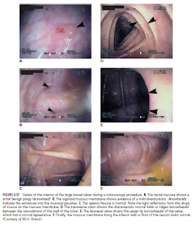
Direct inspection of the lining of the entire colon including the cecum has become an important weapon in the early diagnosis of mucosal polyps and large bowel cancer in recent years.

Not only can the colon be observed and suspicious areas photographed for future reference, but also biopsy specimens can be removed for pathologic examination.
For the diagnosis of early cancer, physicians previously relied almost entirely on rectal examination, sigmoidoscopy, and the detection of occult blood in the feces. The disadvantage of colonoscopy is the high cost. Following a regime in which the large bowel is thoroughly washed out, the patient is relaxed under a light anesthetic. The flexible endoscopic tube is introduced through the anus into the anal canal, rectum, and colon. Colonoscopy can also be used in the diagnosis and treatment of ulcerative colitis and Crohn’s disease.

Colostomy
The sigmoid colon is often selected as a site for performing a colostomy in patients with carcinoma of the rectum. Its mobility allows the surgeon to bring out a loop of colon, with its blood supply intact, through a small incision in the left iliac region of the anterior abdominal wall. Its mobility also makes it suitable for implantation of the ureters after surgical removal of the bladder.


Pancreas-Relations-Trauma of the Pancreas-Cancer of the Head of the Pancreas and the Bile Duct-The Pancreatic Tail and Splenectomy-Diagnosis of Pancreatic Disease

Pancreas
The pancreas is both an exocrine and endocrine gland.
 The exocrine portion of the gland function is  producing secretion that contains enzymes capable of hydrolyzing proteins, fats, and carbohydrates. islets of Langerhans  of the gland, the pancreatic islets (The endocrine portion), produces the hormones insulin and glucagon, which play a key role in carbohydrate metabolism.
It is is an elongated structure that lies in the epigastrium and the left upper quadrant. It is soft and lobulated and situated on the posterior abdominal wall behind the peritoneum. It crosses the transpyloric plane. The pancreas is divided into a head, neck, body, and tail.

The head of the pancreas is disc shaped and lies within the concavity of the duodenum. the uncinate process,is  A part of the head extends to the left behind the superior mesenteric vessels .
The neck which connects the head to the body is the constricted portion of the pancreas and. It lies in front of the beginning of the portal vein and the origin of the superior mesenteric artery from the aorta.
The body runs upward and to the left across the midline . It is somewhat triangular in cross section. The tail passes forward in the splenicorenal ligament and comes in contact with the hilum of the spleen

Relations
■■ Anteriorly: From right to left: the transverse colon and the attachment of the transverse mesocolon, the lesser sac, and the stomach
■■ Posteriorly: From right to left: the bile duct, the portal and splenic veins, the inferior vena cava, the aorta, the origin of the superior mesenteric artery, the left psoas muscle, the left suprarenal gland, the left kidney, and the hilum of the spleen

Trauma of the Pancreas
The pancreas is is well protected because it is deeply placed within the abdomen and  protected by the costal margin and the anterior abdominal wall. However, blunt trauma, such as in a sports injury when a sudden blow to the abdomen occurs, can compress and tear the pancreas against the vertebral column. The pancreas is most commonly damaged by gunshot or stab wounds. Damaged pancreatic tissue releases activated pancreatic enzymes that produce the signs and symptoms of acute peritonitis.
Cancer of the Head of the Pancreas and the Bile Duct
cancer of the head of the pancreas often causes obstructive jaundice Because of the close relation of the head of the pancreas to the bile duct,.

The Pancreatic Tail and Splenectomy
The presence of the tail of the pancreas in the splenicorenal ligament sometimes results in its damage during splenectomy. The damaged pancreas releases enzymes that start to digest surrounding tissues, with serious consequences

Diagnosis of Pancreatic Disease
The deep location of the pancreas sometimes gives rise to problems of diagnosis for the following reasons:
■■ Pain from the pancreas is commonly referred to the back.
■■ Because the pancreas lies behind the stomach and transverse colon, disease of the gland can be confused with that of the stomach or transverse colon.
■■ Inflammation of the pancreas can spread to the peritoneum forming the posterior wall of the lesser sac. This in turn can lead to adhesions and the closing off of the lesser sac to form a pseudocyst.


Friday, 17 June 2016

Colonoscopy-Cancer of the Large Bowel-Cecostomy and Colostomy-Diverticulosis

Colonoscopy
 colonoscopy is now being extensively used for early detection of malignant tumors Since colorectal cancer is a leading cause of death. In this procedure, the mucous membrane of the colon can be directly visualized through an elongated flexible tube, or endoscope. Following a thorough washing out of the large bowel, the patient is sedated, and the tube is gently inserted into the anal canal. The interior of the large bowel can be observed from the anus to the cecum. Photographs of suspicious areas, such as polyps, can be taken and biopsy specimens can be removed for pathologic examination. Although a relatively expensive procedure, it provides a more complete screening examination for colorectal cancer than combined fecal occult blood testing and the examination of the distal colon with sigmoidoscopy .

 
Cancer of the Large Bowel
Cancer of the large bowel is relatively common in persons older than 50 years. The growth is restricted to the bowel wall for a considerable time before it spreads via the lymphatics Bloodstream spread via the portal circulation to the liver occurs late. If a diagnosis is made early and a partial colectomy is performed, accompanied by removal of the lymph vessels and lymph nodes draining the area, then a cure can be anticipated


Cecostomy and Colostomy
Because the cecum, transverse colon, and sigmoid colon, they may be brought to the surface through a small opening in the anterior abdominal wall. If the cecum or transverse colon is then opened, the bowel contents may be allowed to drain by this route. These procedures are referred to as cecostomy or colostomy, respectively, and are used to relieve large-bowel obstructions

Diverticulosis
The term diverticulitis refers to the inflammation of a diverticulum or diverticula, and this may result in perforation of the gut wall Diverticulosis of the colon is a common clinical condition. It consists of a herniation of the lining mucosa through the circular muscle between the teniae coli and occurs at points where the circular muscle is weakest—that is, where the blood vessels pierce the muscle.