Showing posts with label Surgical. Show all posts

Sunday, 26 June 2016

Humerus-Fractures of the Proximal End of the Humerus-Humeral Head Fractures-Greater Tuberosity Fractures-Lesser Tuberosity Fractures-Surgical Neck Fractures-Fractures of the Shaft of the Humerus-Fractures of the Distal End of the Humerus-

Humerus
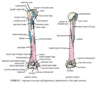
The humerus articulates with the scapula at the shoulder joint and with the radius and ulna at the elbow joint. The upper end of the humerus has a head, which forms about one third of a sphere and articulates with the glenoid cavity of the scapula. Immediately below the head is the anatomic neck. Below the neck are the greater and lesser tuberosities, separated from each other by the bicipital groove. Where the upper end of the humerus joins the shaft is a narrow surgical neck. About halfway down the lateral aspect of the shaft is a roughened elevation called the deltoid tuberosity. Behind and below the tuberosity is a spiral groove, which accommodates the radial nerve The lower end of the humerus possesses the medial and lateral epicondyles for the attachment of muscles and ligaments, the rounded capitulum for articulation with the head of the radius, and the pulley-shaped trochlea for articulation with the trochlear notch of the ulna. Above the capitulum is the radial fossa, which receives the head of the radius when the elbow is flexed. Above the trochlea anteriorly is the coronoid fossa, which during the same movement receives the coronoid process of the ulna. Above the trochlea posteriorly is the olecranon fossa, which receives the olecranon process of the ulna when the elbow joint is extended

Fractures of the Proximal End of the Humerus
Humeral Head Fractures
Fractures of the humeral head can occur during the process of anterior and posterior dislocations of the shoulder joint. The fibrocartilaginous glenoid labrum of the scapula produces the fracture, and the labrum can become jammed in the defect, making reduction of the shoulder joint difficult
Greater Tuberosity Fractures
The greater tuberosity of the humerus can be fractured by direct trauma, displaced by the glenoid labrum during dislocation of the shoulder joint, or avulsed by violent contractions of the supraspinatus muscle. The bone fragment will have the attachments of the supraspinatus, teres minor, and infraspinatus muscles, whose tendons form part of the rotator cuff. When associated with a shoulder dislocation, severe tearing of the cuff with the fracture can result in the greater tuberosity remaining displaced posteriorly after the shoulder joint has been reduced. In this situation, open reduction of the fracture is necessary to attach the rotator cuff back into place.

Lesser Tuberosity Fractures
Occasionally, a lesser tuberosity fracture accompanies posterior dislocation of the shoulder joint. The bone fragment receives the insertion of the subscapularis tendon, a part of the rotator cuff.

Surgical Neck Fractures
The surgical neck of the humerus , which lies immediately distal to the lesser tuberosity, can be fractured by a direct blow on the lateral aspect of the shoulder or in an indirect manner by falling on the outstretched hand.

Fractures of the Shaft of the Humerus
Fractures of the humeral shaft are common; displacement of the fragments depends on the relation of the site of fracture to the insertion of the deltoid muscle. When the fracture line is proximal to the deltoid insertion, the proximal fragment is adducted by the pectoralis major, latissimus dorsi, and teres major muscles; the distal fragment is pulled proximally by the deltoid, biceps, and triceps. When the fracture is distal to the deltoid insertion, the proximal fragment is abducted by the deltoid, and the distal fragment is pulled proximally by the biceps and triceps. The radial nerve can be damaged where it lies in the spiral groove on the posterior surface of the humerus under cover of the triceps muscle.


Fractures of the Distal End of the Humerus
Supracondylar fractures are common in children and occur when the child falls on the outstretched hand with the elbow partially flexed. Injuries to the median, radial, and ulnar nerves are not uncommon, although function usually quickly returns after reduction of the fracture. Damage to or pressure on the brachial artery can occur at the time of the fracture or from swelling of the surrounding tissues; the circulation to the forearm may be interfered with, leading to Volkmann’s ischemic contractureThe medial epicondyle (Fig. 9.10) can be avulsed by themedial collateral ligament of the elbow joint if the forearm is forcibly abducted. The ulnar nerve can be injured at the time of the fracture, can become involved later in the repair process of the fracture (in the callus), or can undergo irritation
on the irregular bony surface after the bone fragments are
reunited
.

  .

Sunday, 19 June 2016

Cushing’s Syndrome-Addison’s Disease-Pheochromocytoma-Surgical Significance of the Renal Fascia-Aortic Aneurysms-Embolic Blockage of the Abdominal Aorta-

Cushing’s Syndrome
Suprarenal cortical hyperplasia is the most common cause of Cushing’s syndrome, the clinical manifestations of which include moon-shaped face, truncal obesity, abnormal hairiness (hirsutism), and hypertension; if the syndrome occurs later in life, it may result from an adenoma or carcinoma of the cortex.

Addison’s Disease
Adrenocortical insufficiency (Addison’s disease), which is characterized clinically by increased pigmentation, muscular weakness, weight loss, and hypotension, may be caused by tuberculous destruction or bilateral atrophy of both cortices.

Pheochromocytoma
Pheochromocytoma, a tumor of the medulla, produces a paroxysmal or sustained hypertension. The symptoms and signs result from the production of a large amount of catecholamines, which are then poured into the bloodstream. Because of their position on the posterior abdominal wall, few tumors of the suprarenal glands can be palpated. CT scans can be used to visualize the glandular enlargement; however, when interpreting CT scans, remember the close relationship of the suprarenal glands to the crura of the diaphragm.

Surgical Significance of the Renal Fascia
The suprarenal glands, together with the kidneys, are enclosed within the renal fascia; the suprarenal glands, however, lie in a separate compartment, which allows the two organs to be separated easily at operation

Aortic Aneurysms
Localized or diffuse dilatations of the abdominal part of the aorta (aneurysms) usually occur below the origin of the renal arteries. Most result from atherosclerosis, which causes weakening of the arterial wall, and occur most commonly in elderly men. Large aneurysms should be treated by open surgical repair. Endovascular repair can also be used by the introduction of a stent graft through one of the iliac arteries with access through the femoral arteries in the groin.

Embolic Blockage of the Abdominal Aorta
The bifurcation of the abdominal aorta where the lumen suddenly narrows may be a lodging site for an embolus discharged from the heart. Severe ischemia of the lower limbs results


Wednesday, 15 June 2016

General Appearances of the Abdominal Wall-Infection of the Umbilicus-Surgical Incisions

General Appearances of the Abdominal Wall
The normal abdominal wall is soft and pliable and undergoes inward and outward excursion with respiration. The contour is subject to considerable variation and depends on the tone of its muscles and the amount of fat in the subcutaneous tissue. Well-developed muscles or an abundance of fat can prove to be a severe obstacle to the palpation of the abdominal viscera
.


Infection of the Umbilicus
In the adult, the umbilicus often receives scant attention in the shower and is consequently a common site of infection.

Surgical Incisions
If possible, all surgical incisions should be made in the lines of cleavage where the bundles of collagen fibers in the dermis run in parallel rows. An incision along a cleavage line will heal as a narrow scar, whereas one that crosses the lines will heal as wide or heaped-up scars.


Tuesday, 14 June 2016

Physical Examination of the Lungs- Trauma to the Lungs-Surgical Access to the Lungs

Physical Examination of the Lungs
For physical examination of the patient, it is helpful to remember that the upper lobes of the lungs are most easily examined from the front of the chest and the lower lobes from the back. In the axillae, areas of all lobes can be examined

Trauma to the Lungs
A physician must always remember that the apex of the lung projects up into the neck (1 in. [2.5 cm] above the clavicle) and can be damaged by stab or bullet wounds in this area. Although the lungs are well protected by the bony thoracic cage, a splinter from a fractured rib can nevertheless penetrate the lung, and air can escape into the pleural cavity, causing a pneumothorax and collapse of the lung. It can also find its way into the lung connective tissue. From there, the air moves under the visceral pleura until it reaches the lung root. It then passes into the mediastinum and up to the neck. Here, it may distend the subcutaneous tissue, a condition known as subcutaneous emphysema. The changes in the position of the thoracic and upper abdominal viscera and the level of the diaphragm during different phases of respiration relative to the chest wall are of considerable clinical importance. A penetrating wound in the lower part of the chest may or may not damage abdominal viscera, depending on the phase of respiration at the time of injury.

Surgical Access to the Lungs
Surgical access to the lung or mediastinum is commonly undertaken through an intercostal space , Special rib retractors that allow the ribs to be widely separated are used. The costal cartilages are sufficiently elastic to permit considerable bending. Good exposure of the lungs is obtained by this method.