Showing posts with label Renal. Show all posts

Sunday, 19 June 2016

Cushing’s Syndrome-Addison’s Disease-Pheochromocytoma-Surgical Significance of the Renal Fascia-Aortic Aneurysms-Embolic Blockage of the Abdominal Aorta-

Cushing’s Syndrome
Suprarenal cortical hyperplasia is the most common cause of Cushing’s syndrome, the clinical manifestations of which include moon-shaped face, truncal obesity, abnormal hairiness (hirsutism), and hypertension; if the syndrome occurs later in life, it may result from an adenoma or carcinoma of the cortex.

Addison’s Disease
Adrenocortical insufficiency (Addison’s disease), which is characterized clinically by increased pigmentation, muscular weakness, weight loss, and hypotension, may be caused by tuberculous destruction or bilateral atrophy of both cortices.

Pheochromocytoma
Pheochromocytoma, a tumor of the medulla, produces a paroxysmal or sustained hypertension. The symptoms and signs result from the production of a large amount of catecholamines, which are then poured into the bloodstream. Because of their position on the posterior abdominal wall, few tumors of the suprarenal glands can be palpated. CT scans can be used to visualize the glandular enlargement; however, when interpreting CT scans, remember the close relationship of the suprarenal glands to the crura of the diaphragm.

Surgical Significance of the Renal Fascia
The suprarenal glands, together with the kidneys, are enclosed within the renal fascia; the suprarenal glands, however, lie in a separate compartment, which allows the two organs to be separated easily at operation

Aortic Aneurysms
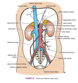
Localized or diffuse dilatations of the abdominal part of the aorta (aneurysms) usually occur below the origin of the renal arteries. Most result from atherosclerosis, which causes weakening of the arterial wall, and occur most commonly in elderly men. Large aneurysms should be treated by open surgical repair. Endovascular repair can also be used by the introduction of a stent graft through one of the iliac arteries with access through the femoral arteries in the groin.

Embolic Blockage of the Abdominal Aorta
The bifurcation of the abdominal aorta where the lumen suddenly narrows may be a lodging site for an embolus discharged from the heart. Severe ischemia of the lower limbs results


Ureter- Ureteric Stones-Renal Colic-Traumatic Ureteral Injuries

Ureter
Each ureter measures about 10 in. (25 cm) long,having three constrictions along its course where the renal pelvis joins the ureter, where it is kinked as it crosses the pelvic brim, and where it pierces the bladder wall


Ureteric Stones
There are three sites of anatomic narrowing of the ureter where stones may be arrested, namely, the pelviureteral junction, the pelvic brim, and where the ureter enters the bladder. Most stones, although radiopaque, are small enough to be impossible to see definitely along the course of the ureter on plain radiographic examination. An intravenous pyelogram is usually necessary. The ureter runs down in front of the tips of the transverse processes of the lumbar vertebrae, crosses the region of the sacroiliac joint, swings out to the ischial spine, and then turns medially to the bladder

Renal Colic
The renal pelvis and the ureter send their afferent nerves into the spinal cord at segments T11 and 12 and L1 and 2. In renal colic, strong peristaltic waves of contraction pass down the ureter in an attempt to pass the stone onward. The spasm of the smooth muscle causes an agonizing colicky pain, which is referred to the skin areas that are supplied by these segments of the spinal cord, namely, the flank, loin, and groin.
When a stone enters the low part of the ureter, the pain is felt at a lower level and is often referred to the testis or the tip of the penis in the male and the labium majus in the female. Sometimes, ureteral pain is referred along the femoral branch of the genitofemoral nerve (L1 and 2) so that pain is experienced in the front of the thigh. The pain is often so severe that afferent pain impulses spread within the central nervous system, giving rise to nausea.

Traumatic Ureteral Injuries
Because of its protected position and small size, injuries to the ureter are rare. Most injuries are caused by gunshot wounds and, in a few individuals, penetrating stab wounds. Because the ureters are retroperitoneal in position, urine may escape into the retroperitoneal tissues on the posterior abdominal wall.


Kidneys-Renal Mobility-Kidney Trauma-Kidney Tumors-Renal Pain-Transplanted Kidneys-

Kidneys
The kidneys function is to excrete most of the waste products of metabolism. alsoThey play a major role in controlling the water and electrolyte balance within the body and in maintaining the acid–base balance of the blood. The waste products leave the kidneys as urine, which passes down the ureters to the urinary bladder, located within the pelvis. The urine leaves the body in the urethra.
The kidneys are reddish brown and lie behind the peritoneum high up on the posterior abdominal wall on either side of the vertebral column; they are largely under cover of the costal margin.
The right kidney lies slightly lower than the left kidney because of the large size of the right lobe of the liver. With contraction of the diaphragm during respiration, both kidneys move downward in a vertical direction by as much as 1 in. (2.5 cm). On the medial concave border of each kidney is a vertical slit that is bounded by thick lips of renal substance and is called the hilum. The hilum extends into a large cavity called the renal sinus. The hilum transmits, from the front backward, the renal vein, two branches of the renal artery, the ureter, and the third branch of the renal artery (VAUA). Lymph vessels and sympathetic fibers also pass through the hilum

Renal Mobility
The kidneys are maintained in their normal position by intraabdominal pressure and by their connections with the perirenal fat and renal fascia. Each kidney moves slightly with respiration. The right kidney lies at a slightly lower level than the left kidney, and the lower pole may be palpated in the right lumbar region at the end of deep inspiration in a person with poorly developed abdominal musculature. Should the amount of perirenal fat be reduced, the mobility of the kidney may become excessive and produce symptoms of renal colic caused by kinking of the ureter. Excessive mobility of the kidney leaves the suprarenal gland undisturbed because the latter occupies a separate compartment in the renal fascia.


Kidney Trauma
The kidneys are well protected by the lower ribs, the lumbar muscles, and the vertebral column. However, a severe blunt injury applied to the abdomen may crush the kidney against the last rib and the vertebral column. Depending on the severity of the blow, the injury varies from a mild bruising to a complete laceration of the organ. Penetrating injuries are usually caused by stab wounds or gunshot wounds and often involve other viscera. Because 25% of the cardiac outflow passes through the kidneys, renal injury can result in rapid blood loss


Kidney Tumors
Malignant tumors of the kidney have a strong tendency to spread along the renal vein. The left renal vein receives the left testicular vein in the male, and this may rarely become blocked, producing left-sided varicocele.

Renal Pain
Renal pain varies from a dull ache to a severe pain in the flank that may radiate downward into the lower abdomen. Renal pain can result from stretching of the kidney capsule or spasm of the smooth muscle in the renal pelvis. The afferent nerve fibers pass through the renal plexus around the renal artery and ascend to the spinal cord through the lowest splanchnic nerve in the thorax and the sympathetic trunk. They enter the spinal cord at the level of T12. Pain is commonly referred along the distribution of the subcostal nerve (T12) to the flank and the anterior abdominal wall.

Transplanted Kidneys
The iliac fossa on the posterior abdominal wall is the usual site chosen for transplantation of the kidney. The fossa is exposed through an incision in the anterior abdominal wall just above the inguinal ligament. The iliac fossa in front of the iliacus muscle is approached retroperitoneally. The kidney is positioned and the vascular anastomosis constructed. The renal artery is anastomosed end to end to the internal iliac artery and the renal vein is anastomosed end to side to the external iliac vein. Anastomosis of the branches of the internal iliac arteries on the two sides is sufficient so that the pelvic viscera on the side of the renal arterial anastomosis are not at risk. Ureterocystostomy is then performed by opening the bladder and providing a wide entrance of the ureter through the bladder wall.