Showing posts with label Vaginal. Show all posts

Friday, 24 June 2016

Male Urogenital Triangle-Penis-Scrotum-Testes-Epididymides-Female Urogenital Triangle-Vulva-Mons Pubis-Labia Majora-Labia Minora-Vestibule-Vaginal Orifice-Clitoris

Male Urogenital Triangle
The male urogenital triangle contains the penis and the scrotum.

Penis
The penis consists of a root, a body, and a glans. The root of the penis consists of three masses of erectile tissue called the bulb of the penis and the right and left crura of the penis. The bulb can be felt on deep palpation in the midline of the perineum, posterior to the scrotum.
The body of the penis is the free portion of the penis, which is suspended from the symphysis pubis. Note that the dorsal surface (anterior surface of the flaccid organ) usually possesses a superficial dorsal vein in the midline The glans penis forms the extremity of the body of the penis. At the summit of the glans is the external urethral meatus. Extending from the lower margin of the external meatus is a fold connecting the glans to the prepuce called the frenulum. The edge of the base of the glans is called the corona. The prepuce or foreskin is formed by a fold of skin attached to the neck of the penis. The prepuce covers the glans for a variable extent, and it should be possible to retract it over the glans.


Scrotum
The scrotum is a sac of skin and fascia containing the testes and the epididymides. The skin of the scrotum is rugose and is covered with sparse hairs. The bilateral origin of the scrotum is indicated by the presence of a dark line in the midline, called the scrotal raphe, along the line of fusion
.
Testes
The testes should be palpated. They are oval shaped and have a firm consistency. They lie free within the tunica vaginalis and are not tethered to the subcutaneous tissue or skin.

Epididymides
Each epididymis can be palpated on the posterolateral surface of the testis. The epididymis is a long, narrow, firm structure having an expanded upper end or head, a body, and a pointed tail inferiorly. The cordlike vas deferens emerges from the tail and ascends medial to the epididymis to enter the spermatic cord at the upper end of the scrotum.


Female Urogenital Triangle
Vulva
“Vulva” is the term applied to the female external genitalia

Mons Pubis
The mons pubis is the rounded, hair-bearing elevation of skin found anterior to the pubis. The pubic hair in the female has an abrupt horizontal superior margin, whereas in the male it extends upward to the umbilicus.

Labia Majora
The labia majora are prominent, hair-bearing folds of skin extending posteriorly from the mons pubis to unite posteriorly in the midline
.
Labia Minora
The labia minora are two smaller, hairless folds of soft skin that lie between the labia majora . Their posterior ends are united to form a sharp fold, the fourchette. Anteriorly, they split to enclose the clitoris, forming an anterior prepuce and a posterior frenulum

Vestibule
The vestibule is a smooth triangular area bounded laterally by the labia minora, with the clitoris at its apex and the fourchette at its base.

Vaginal Orifice
The vaginal orifice is protected in virgins by a thin mucosal fold called the hymen, which is perforated at its center. At the first coitus, the hymen tears, usually posteriorly or posterolaterally, and after childbirth only a few tags of the hymen remain .

Clitoris
This is situated at the apex of the vestibule anteriorly. The glans of the clitoris is partly hidden by the prepuce.


Vaginal Examination-Injury to the Perineum during Childbirth-Pudendal Nerve Block-Area of Anesthesia-Indications-Transvaginal Procedure-Perineal Procedure

Vaginal Examination
Digital examination of the vagina may provide the physician with much valuable information concerning the health of the vaginal walls, the uterus, and the surrounding structures
Injury to the Perineum during Childbirth
The perineal body is a wedge of fibromuscular tissue that lies between the lower part of the vagina and the anal canal. It is held in position by the insertion of the perineal muscles and by the attachment of the levator ani muscles. In the female, it is a much larger structure than in the male, and it serves to support the posterior wall of the vagina. Damage by laceration during childbirth can be followed by permanent weakness of the pelvic floor.

 Few women escape some injury to the birth canal during delivery. In most, this is little more than an abrasion of the posterior vaginal wall. Spontaneous delivery of the child with the patient unattended can result in a severe tear of the lower third of the posterior wall of the vagina, the perineal body, and overlying skin. In severe tears, the lacerations may extend backward into the anal canal and damage the external sphincter. In these cases, it is imperative that an accurate repair of the walls of the anal canal, vagina, and perineal body be undertaken as soon as possible.

In the management of childbirth, when it is obvious to the obstetrician that the perineum will tear before the baby’s head emerges through the vaginal orifice, a planned surgical incision is made through the perineal skin in a posterolateral direction to avoid the anal sphincters. This procedure is known as an episiotomy. Breech deliveries and forceps deliveries are usually preceded by an episiotomy
.
Pudendal Nerve Block
Area of Anesthesia
The area anesthetized is the skin of the perineum; this nerve block does not, however, abolish sensation from the anterior part of the perineum, which is innervated by the ilioinguinal nerve and the genitofemoral nerve. Needless to say, it does not abolish pain from uterine contractions that ascend to the spinal cord via the sympathetic afferent nerves.


Indications
During the second stage of a difficult labor, when the presenting part of the fetus, usually the head, is descending through the vulva, forceps delivery and episiotomy may be necessary
.
Transvaginal Procedure
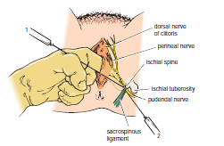
The bony landmark used is the ischial spine . The index finger is inserted through the vagina to palpate the ischial spine. The needle of the syringe is then passed through the vaginal mucous membrane toward the ischial spine. On passing through the sacrospinous ligament, the anesthetic solution is injected around the pudendal nerve .

Perineal Procedure
The bony landmark is the ischial tuberosity . The tuberosity is palpated subcutaneously through the buttock, and the needle is introduced into the pudendal canal along the medial side of the tuberosity. The canal lies about 1 in. (2.5 cm) deep to the free surface of the ischial tuberosity. The local anesthetic is then infiltrated around the pudendal nerve.


Vagina-Relations-Function-Supports of the Vagina-Vaginal Examination-Prolapse of the Vagina-Vaginal Trauma

Vagina
The vagina is a muscular tube that extends upward and backward from the vulva to the uterus. It measures about 3 in. (8 cm) long and has anterior and posterior walls, which are normally in apposition. At its upper end, the anterior wall is pierced by the cervix, which projects downward and backward into the vagina. It is important to remember that the upper half of the vagina lies above the pelvic floor and the lower half lies within the perineum. The area of the vaginal lumen, which surrounds the cervix, is divided into four regions, or fornices: anterior, posterior, right lateral, and left lateral. The vaginal orifice in a virgin possesses a thin mucosal fold called the hymen, which is perforated at its center. After childbirth, the hymen usually consists only of tags

Relations
■■ Anteriorly: The vagina is closely related to the bladder above and to the urethra below.
■■ Posteriorly: The upper third of the vagina is related to the rectouterine pouch (pouch of Douglas) and its middle third to the ampulla of the rectum. The lower third is related to the perineal body, which separates it from the anal canal.

■■ Laterally: In its upper part, the vagina is related to the ureter; its middle part is related to the anterior fibers of the levator ani, as they run backward to reach the perineal body and hook around the anorectal junction. Contraction of the fibers of levator ani compresses the walls of the vagina together. In its lower part, the vagina is related to the urogenital diaphragm and the bulb of the vestibule.

 
Function
The vagina not only is the female genital canal, but it also serves as the excretory duct for the menstrual flow and

Supports of the Vagina
The upper part of the vagina is supported by the levatores ani muscles and the transverse cervical, pubocervical, and sacrocervical ligaments. These structures are attached to the vaginal wall by pelvic fascia. The middle part of the vagina is supported by the urogenital diaphragm. The lower part of the vagina, especially the posterior wall, is supported by the perineal body.

Vaginal Examination
The anatomic relations of the vagina are of great clinical importance. Many pathologic conditions occurring in the female pelvis may be diagnosed using a simple vaginal examination.
The following structures can be palpated through the vaginal walls from above downward:
■■ Anteriorly: The bladder and the urethra
■■ Posteriorly: Loops of ileum and the sigmoid colon in the rectouterine peritoneal pouch (pouch of Douglas), the rectal ampulla, and the perineal body
■■ Laterally: The ureters, the pelvic fascia and the anterior fibers of the levatores ani muscles, and the urogenital diaphragm

Prolapse of the Vagina
The vaginal vault is supported by the same structures that support the uterine cervix. Prolapse of the uterus is necessarily associated with some degree of sagging of the vaginal walls. However, if the supports of the bladder, urethra, or anterior rectal wall are damaged in childbirth, prolapse of the vaginal walls occurs, with the uterus remaining in its correct position.
Sagging of the bladder results in the bulging of the anterior wall of the vagina, a condition known as a cystocele. When the ampulla of the rectum sags against the posterior vaginal wall, the bulge is called a rectocele.

Vaginal Trauma
Coital injury, picket fence–type of impalement injury, and vaginal perforation caused by water under pressure, as occurs in water skiing, are common injuries. Lacerations of the vaginal wall involving the posterior fornix may violate the pouch of Douglas of the peritoneal cavity and cause prolapse of the small intestine into the vagina

forms part of the birth canal.