Showing posts with label Malignant. Show all posts

Sunday, 19 June 2016

Sacral Plexus-Pressure from the Fetal Head-Invasion by Malignant Tumors-Referred Pain from the Obturator Nerve-Caudal Anesthesia (Analgesia)

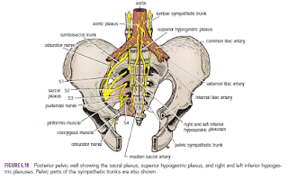
Sacral Plexus
The sacral plexus lies in front of the piriformis muscle on the posterior pelvic wall. It is formed from the anterior rami of the 4th and 5th lumbar nerves and the anterior rami of the first, second, third, and fourth sacral nerves. The fourth lumbar nerve joins the fifth lumbar nerve to form the lumbosacral trunk. The lumbosacral trunk passes down into the pelvis and joins the sacral nerves as they emerge from the anterior sacral foramina.

 
Pressure from the Fetal Head
when the fetal head has descended into the pelvis During the later stages of pregnancy, , the mother often complains of discomfort or aching pain extending down one of the lower limbs. The discomfort, caused by pressure from the fetal head, is often relieved by changing position, such as lying on the side in bed.

Invasion by Malignant Tumors
The nerves of the sacral plexus can become invaded by malignant tumors extending from neighboring viscera. A carcinoma of the rectum, for example, can cause severe intractable pain down the lower limbs.

Referred Pain from the Obturator Nerve
The obturator nerve lies on the lateral wall of the pelvis and supplies the parietal peritoneum. An inflamed appendix hanging down into the pelvic cavity could cause irritation of the obturator nerve endings, leading to referred pain down the inner side of the right thigh. Inflammation of the ovaries can produce similar symptoms.

Caudal Anesthesia (Analgesia)
Anesthetic solutions can be injected into the sacral canal through the sacral hiatus. The solutions then act on the spinal roots of the 2nd, 3rd, 4th and 5th sacral and coccygeal segments of the cord as they emerge from the dura mater. The roots of higher spinal segments can also be blocked by this method. The needle must be confined to the lower part of the sacral canal, because the meninges extend down as far as the lower border of the second sacral vertebra. Caudal anesthesia is used in obstetrics to block pain fibers from the cervix of the uterus and to anesthetize the perineum


Friday, 17 June 2016

Portal–Systemic Anastomoses-Portal Hypertension-Blood Flow in the Portal Vein and Malignant Disease-

Portal–Systemic Anastomoses
Under normal conditions, the portal venous blood traverses the liver and drains into the inferior vena cava of the systemic venous circulation by way of the hepatic veins. This is the direct route. However, other, smaller communications exist between the portal and systemic systems, and they become important when the direct route becomes blocked

These communications are as follows
:
■■ At the lower third of the esophagus, the esophageal branches of the left gastric vein (portal tributary) anastomose with the esophageal veins draining the middle third of the esophagus into the azygos veins (systemic tributary).

■■ Halfway down the anal canal, the superior rectal veins (portal tributary) draining the upper half of the anal canal anastomose with the middle and inferior rectal veins (systemic tributaries), which are tributaries of the internal iliac and internal pudendal veins, respectively.

■■ The paraumbilical veins connect the left branch of the portal vein with the superficial veins of the anterior abdominal wall (systemic tributaries). The paraumbilical veins travel in the falciform ligament and accompany the ligamentum teres.


■■ The veins of the ascending colon, descending colon, duodenum, pancreas, and liver (portal tributary) anastomose with the renal, lumbar, and phrenic veins (systemic tributaries).

Portal Hypertension
Portal hypertension is a common clinical condition; thus, the list of portal–systemic anastomoses should be remembered. Enlargement of the portal–systemic connections is frequently accompanied by congestive enlargement of the spleen. Portacaval shunts for the treatment of portal hypertension may involve the anastomosis of the portal vein, because it lies within the lesser omentum, to the anterior wall of the inferior vena cava behind the entrance into the lesser sac. The splenic vein may be anastomosed to the left renal vein after removing the spleen.


Blood Flow in the Portal Vein and Malignant Disease
The portal vein conveys about 70% of the blood to the liver. The remaining 30% is oxygenated blood, which passes to the liver via the hepatic artery. The wide angle of union of the splenic vein with the superior mesenteric vein to form the portal vein leads to streaming of the blood flow in the portal vein. The right lobe of the liver receives blood mainly from the intestine, whereas the left lobe plus the quadrate and caudate lobes receive blood from the stomach and the spleen. This distribution of blood may explain the distribution of secondary malignant deposits in the liver.


Wednesday, 15 June 2016

Varicocele-Malignant Tumor of the Testis-Torsion of the Testis-Processus Vaginalis

Varicocele
A varicocele is a condition in which the veins of the pampiniform plexus are elongated and dilated. It is a common disorder in adolescents and young adults, with most occurring on the left side. This is thought to be because the right testicular vein joins the low-pressure inferior vena cava, whereas the left vein joins the left renal vein, in which the venous pressure is higher. Rarely, malignant disease of the left kidney extends along the renal vein and blocks the exit of the testicular vein. A rapidly developing left-sided variocele should therefore always lead one to examine the left kidney.

 
Malignant Tumor of the Testis
A malignant tumor of the testis spreads upward via the lymph vessels to the lumbar (para-aortic) lymph nodes at the level of the first lumbar vertebra. It is only later, when the tumor spreads locally to involve the tissues and skin of the scrotum, that the superficial inguinal lymph nodes are involved. The testis may be subject to the following congenital anomalies.

Torsion of the Testis
Torsion of the testis is a rotation of the testis around the spermatic cord within the scrotum. It is often associated with an excessively large tunica vaginalis. Torsion commonly occurs in active young men and children and is accompanied by severe pain. If not treated quickly, the testicular artery may be occluded, followed by necrosis of the testis
.

Processus Vaginalis
The formation of the processus vaginalis and its passage through the lower part of the anterior abdominal wall with the formation of the inguinal canal in both sexes were described elsewhere . Normally, the upper part becomes obliterated just before birth and the lower part remains as the tunica vaginalis.