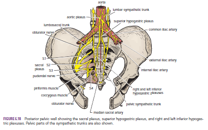
Showing posts with label from. Show all posts

Sunday, 19 June 2016

Sacral Plexus-Pressure from the Fetal Head-Invasion by Malignant Tumors-Referred Pain from the Obturator Nerve-Caudal Anesthesia (Analgesia)

Sacral Plexus
The sacral plexus lies in front of the piriformis muscle on the posterior pelvic wall. It is formed from the anterior rami of the 4th and 5th lumbar nerves and the anterior rami of the first, second, third, and fourth sacral nerves. The fourth lumbar nerve joins the fifth lumbar nerve to form the lumbosacral trunk. The lumbosacral trunk passes down into the pelvis and joins the sacral nerves as they emerge from the anterior sacral foramina.

 
Pressure from the Fetal Head
when the fetal head has descended into the pelvis During the later stages of pregnancy, , the mother often complains of discomfort or aching pain extending down one of the lower limbs. The discomfort, caused by pressure from the fetal head, is often relieved by changing position, such as lying on the side in bed.

Invasion by Malignant Tumors
The nerves of the sacral plexus can become invaded by malignant tumors extending from neighboring viscera. A carcinoma of the rectum, for example, can cause severe intractable pain down the lower limbs.

Referred Pain from the Obturator Nerve
The obturator nerve lies on the lateral wall of the pelvis and supplies the parietal peritoneum. An inflamed appendix hanging down into the pelvic cavity could cause irritation of the obturator nerve endings, leading to referred pain down the inner side of the right thigh. Inflammation of the ovaries can produce similar symptoms.

Caudal Anesthesia (Analgesia)
Anesthetic solutions can be injected into the sacral canal through the sacral hiatus. The solutions then act on the spinal roots of the 2nd, 3rd, 4th and 5th sacral and coccygeal segments of the cord as they emerge from the dura mater. The roots of higher spinal segments can also be blocked by this method. The needle must be confined to the lower part of the sacral canal, because the meninges extend down as far as the lower border of the second sacral vertebra. Caudal anesthesia is used in obstetrics to block pain fibers from the cervix of the uterus and to anesthetize the perineum


Saturday, 11 June 2016

Sino-atrial Block-Atrio-ventricular (A-V) Block- Incomplete A-V Block -

Arrhythmia Resulting from Conductive Conductive Block
Sino-atrial Block

In rare instances, SAN impulse is blocked before it enters the atrial muscle.
In ECG there is sudden cessation of P waves, with resultant standstill of the atria.
The ventricles pick up a new spontaneous rhythm, usually originating from the A-V node.
So the rate of the ventricular QRS-T complex is slowed down
Otherwise, ventricular depolarization (QRS) and repolarization (T) waves are not altered

Atrio-ventricular (A-V) Block
Conditions that can slow impulse conduction rate in bundle of His or block the impulse entirely are:
Ischemia of A-V node or A-V bundle fibers resulting from coronary insufficiency.
Compression of the A-V bundle by scar tissue or calcified portions of the heart.
Inflammation of A-V node or A-V bundle (resulting from different types of myocarditis, (diphtheria / rheumatic fever).
Extreme stimulation of the heart by the vagus nerves in rare instances blocks impulse conduction through the A-V node.

Incomplete A-V Block
Prolonged P-R Interval (First-Degree Block
)
The usual lapse of time between beginning of the P wave and beginning of the QRS complex (P-R interval) is about 0.16 sec. 
When P-R interval > 0.20 sec, it is said to be prolonged & the patient is said to have first-degree incomplete heart block.
Thus, first-degree block is defined as a delay of conduction from the atria to the ventricles but not actual blockage of conduction.
P-R interval seldom increases > 0.35-0.45 sec, by that time, conduction through A-V bundle stops entirely.
One means for determining severity of some heart diseases (acute rheumatic heart disease) is to measure P-R interval


.
Incomplete A-V Block
(Second-Degree Block)
When A-V bundle conduction is significantly slowed, sometimes AP is strong enough (& sometimes is not) to pass through the bundle.
In this instance, there will be atrial P wave but no QRS-T wave, & it is said that there are "dropped beats" of the ventricles.
This condition is called second-degree heart block.
At times, every other beat of the ventricles is dropped, so a "2:1 rhythm" develops.
At other times, rhythms of 3:2 or 3:1 also develop.