Showing posts with label Tumors. Show all posts

Sunday, 19 June 2016

Sacral Plexus-Pressure from the Fetal Head-Invasion by Malignant Tumors-Referred Pain from the Obturator Nerve-Caudal Anesthesia (Analgesia)

Sacral Plexus
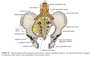
The sacral plexus lies in front of the piriformis muscle on the posterior pelvic wall. It is formed from the anterior rami of the 4th and 5th lumbar nerves and the anterior rami of the first, second, third, and fourth sacral nerves. The fourth lumbar nerve joins the fifth lumbar nerve to form the lumbosacral trunk. The lumbosacral trunk passes down into the pelvis and joins the sacral nerves as they emerge from the anterior sacral foramina.

 
Pressure from the Fetal Head
when the fetal head has descended into the pelvis During the later stages of pregnancy, , the mother often complains of discomfort or aching pain extending down one of the lower limbs. The discomfort, caused by pressure from the fetal head, is often relieved by changing position, such as lying on the side in bed.

Invasion by Malignant Tumors
The nerves of the sacral plexus can become invaded by malignant tumors extending from neighboring viscera. A carcinoma of the rectum, for example, can cause severe intractable pain down the lower limbs.

Referred Pain from the Obturator Nerve
The obturator nerve lies on the lateral wall of the pelvis and supplies the parietal peritoneum. An inflamed appendix hanging down into the pelvic cavity could cause irritation of the obturator nerve endings, leading to referred pain down the inner side of the right thigh. Inflammation of the ovaries can produce similar symptoms.

Caudal Anesthesia (Analgesia)
Anesthetic solutions can be injected into the sacral canal through the sacral hiatus. The solutions then act on the spinal roots of the 2nd, 3rd, 4th and 5th sacral and coccygeal segments of the cord as they emerge from the dura mater. The roots of higher spinal segments can also be blocked by this method. The needle must be confined to the lower part of the sacral canal, because the meninges extend down as far as the lower border of the second sacral vertebra. Caudal anesthesia is used in obstetrics to block pain fibers from the cervix of the uterus and to anesthetize the perineum


Kidneys-Renal Mobility-Kidney Trauma-Kidney Tumors-Renal Pain-Transplanted Kidneys-

Kidneys
The kidneys function is to excrete most of the waste products of metabolism. alsoThey play a major role in controlling the water and electrolyte balance within the body and in maintaining the acid–base balance of the blood. The waste products leave the kidneys as urine, which passes down the ureters to the urinary bladder, located within the pelvis. The urine leaves the body in the urethra.
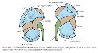
The kidneys are reddish brown and lie behind the peritoneum high up on the posterior abdominal wall on either side of the vertebral column; they are largely under cover of the costal margin.
The right kidney lies slightly lower than the left kidney because of the large size of the right lobe of the liver. With contraction of the diaphragm during respiration, both kidneys move downward in a vertical direction by as much as 1 in. (2.5 cm). On the medial concave border of each kidney is a vertical slit that is bounded by thick lips of renal substance and is called the hilum. The hilum extends into a large cavity called the renal sinus. The hilum transmits, from the front backward, the renal vein, two branches of the renal artery, the ureter, and the third branch of the renal artery (VAUA). Lymph vessels and sympathetic fibers also pass through the hilum

Renal Mobility
The kidneys are maintained in their normal position by intraabdominal pressure and by their connections with the perirenal fat and renal fascia. Each kidney moves slightly with respiration. The right kidney lies at a slightly lower level than the left kidney, and the lower pole may be palpated in the right lumbar region at the end of deep inspiration in a person with poorly developed abdominal musculature. Should the amount of perirenal fat be reduced, the mobility of the kidney may become excessive and produce symptoms of renal colic caused by kinking of the ureter. Excessive mobility of the kidney leaves the suprarenal gland undisturbed because the latter occupies a separate compartment in the renal fascia.


Kidney Trauma
The kidneys are well protected by the lower ribs, the lumbar muscles, and the vertebral column. However, a severe blunt injury applied to the abdomen may crush the kidney against the last rib and the vertebral column. Depending on the severity of the blow, the injury varies from a mild bruising to a complete laceration of the organ. Penetrating injuries are usually caused by stab wounds or gunshot wounds and often involve other viscera. Because 25% of the cardiac outflow passes through the kidneys, renal injury can result in rapid blood loss


Kidney Tumors
Malignant tumors of the kidney have a strong tendency to spread along the renal vein. The left renal vein receives the left testicular vein in the male, and this may rarely become blocked, producing left-sided varicocele.

Renal Pain
Renal pain varies from a dull ache to a severe pain in the flank that may radiate downward into the lower abdomen. Renal pain can result from stretching of the kidney capsule or spasm of the smooth muscle in the renal pelvis. The afferent nerve fibers pass through the renal plexus around the renal artery and ascend to the spinal cord through the lowest splanchnic nerve in the thorax and the sympathetic trunk. They enter the spinal cord at the level of T12. Pain is commonly referred along the distribution of the subcostal nerve (T12) to the flank and the anterior abdominal wall.

Transplanted Kidneys
The iliac fossa on the posterior abdominal wall is the usual site chosen for transplantation of the kidney. The fossa is exposed through an incision in the anterior abdominal wall just above the inguinal ligament. The iliac fossa in front of the iliacus muscle is approached retroperitoneally. The kidney is positioned and the vascular anastomosis constructed. The renal artery is anastomosed end to end to the internal iliac artery and the renal vein is anastomosed end to side to the external iliac vein. Anastomosis of the branches of the internal iliac arteries on the two sides is sufficient so that the pelvic viscera on the side of the renal arterial anastomosis are not at risk. Ureterocystostomy is then performed by opening the bladder and providing a wide entrance of the ureter through the bladder wall.


Tuesday, 14 June 2016

Deflection of Mediastinum-Mediastinitis-Mediastinal Tumors or Cysts-Mediastinoscopy

Deflection of Mediastinum
In the cadaver, the mediastinum, as the result of the hardening effect of the preserving fluids, is an inflexible, fixed structure. In the living, it is very mobile; the lungs, heart, and large arteries are in rhythmic pulsation, and the esophagus distends as each bolus of food passes through it.
If air enters the pleural cavity (a condition called pneumothorax), the lung on that side immediately collapses and the mediastinum is displaced to the opposite side. This condition reveals itself by the patient’s being breathless and in a state of shock; on examination, the trachea and the heart are found to be displaced to the opposite side


Mediastinitis
The structures that make up the mediastinum are embedded in loose connective tissue that is continuous with that of the root of the neck. Thus, it is possible for a deep infection of the neck to spread readily into the thorax, producing a mediastinitis. Penetrating wounds of the chest involving the esophagus may produce a mediastinitis. In esophageal perforations, air escapes into the connective tissue spaces and ascends beneath the fascia to the root of the neck, producing subcutaneous emphysema.

Mediastinal Tumors or Cysts
Because many vital structures are crowded together within the mediastinum, their functions can be interfered with by an enlarging tumor or organ. A tumor of the left lung can rapidly spread to involve the mediastinal lymph nodes, which on enlargement may compress the left recurrent laryngeal nerve, producing paralysis of the left vocal fold. An expanding cyst or tumor can partially occlude the superior vena cava, causing severe congestion of the veins of the upper part of the body. Other pressure effects can be seen on the sympathetic trunks, phrenic nerves, and sometimes the trachea, main bronchi, and esophagus.

Mediastinoscopy
Mediastinoscopy is a diagnostic procedure whereby specimens of tracheobronchial lymph nodes are obtained without opening the pleural cavities. A small incision is made in the midline in the neck just above the suprasternal notch, and the superior mediastinum is explored down to the region of the bifurcation of the trachea. The procedure can be used to determine the diagnosis and degree of spread of carcinoma of the bronchus

.