Wednesday 29 June 2016

Arterial Injury of the upper limb-Palpation and Compression of Arteries-Allen Test-Arterial Innervation and Raynaud’s Disease-

Arterial Injury of the upper limb
The arteries of the upper limb can be damaged by penetrating wounds or may require ligation in amputation operations.
Because of the existence of an adequate collateral circulation around the shoulder, elbow, and wrist joints, ligation of the main arteries of the upper limb is not followed by tissue necrosis or gangrene, provided, of course, that the arteries forming the collateral circulation are not diseased and the patient’s general circulation is satisfactory. Nevertheless, it can take days or weeks for the collateral vessels to open sufficiently to provide the distal part of the limb with the same volume of blood as previously supplied by the main artery.

Palpation and Compression of Arteries
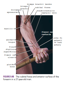
A clinician must know where the arteries of the upper limb can be palpated or compressed in an emergency. The subclavian artery, as it crosses the first rib to become the axillary artery, can be palpated in the root of the posterior triangle of the neck. The artery can be compressed here against the first rib to stop a catastrophic hemorrhage. The third part of the axillary artery can be felt in the axilla as it lies in front of the teres major muscle. The brachial artery can be palpated in the arm as it lies on the brachialis and is overlapped from the lateral side by the biceps brachii.
The radial artery lies superficially in front of the distal end of the radius, between the tendons of the brachioradialis and flexor carpi radialis; it is here that the clinician takes the radial pulse. If the pulse cannot be felt, try feeling for the radial artery on the other wrist; occasionally, a congenitally abnormal radial artery can be difficult to feel. The radial artery can be less easily felt as it crosses the anatomic snuffbox.
The ulnar artery can be palpated as it crosses anterior to the flexor retinaculum in company with the ulnar nerve. The artery lies lateral to the pisiform bone, separated from it by the ulnar nerve. The artery is commonly damaged here in laceration wounds in front of the wrist.

Allen Test
The Allen test is used to determine the patency of the ulnar and radial arteries. With the patient’s hands resting in the lap, compress the radial arteries against the anterior surface of each radius and ask the patient to tightly clench the fists. The clenching of the fists closes off the superficial and deep palmar arterial arches. When the patient is asked to open the hands, the skin of the palms is at first white, and then normally the blood quickly flows into the arches through the ulnar arteries, causing the palms to promptly turn pink. This establishes that the ulnar arteries are patent. The patency of the radial arteries can be established by repeating the test but this time compressing the ulnar arteries as they lie lateral to the pisiform bones.

Arterial Innervation and Raynaud’s Disease
The arteries of the upper limb are innervated by sympathetic nerves. The preganglionic fibers originate from cell bodies in the 2nd to 8th thoracic segments of the spinal cord. They ascend in the sympathetic trunk and synapse in the middle cervical, inferior cervical, 1st thoracic, or stellate ganglia. The postganglionic fibers join the nerves that form the brachial plexus and are distributed to the arteries within the branches of the plexus.
For example, the digital arteries of the fingers are supplied by postganglionic sympathetic fibers that run in the digital nerves. Vasospastic diseases involving digital arterioles, such as Raynaud’s disease, may require a cervicodorsal preganglionic sympathectomy to prevent necrosis of the fingers. The operation is followed by arterial vasodilatation, with consequent increased blood flow to the upper limb.


The Wrist and Hand-Important Structures Lying in Front of the Wrist-Radial Artery-Tendon of Flexor Carpi Radialis-Tendon of Palmaris Longus (If Present)-Tendons of Flexor Digitorum Superficialis-Tendon of Flexor Carpi Ulnaris-Ulnar Artery-Ulnar Nerve-Important Structures Lying on the Lateral Side of the Wrist-Anatomic Snuffbox-Important Structures Lying on the Back of the Wrist-Lunate-Important Structures Lying in the Palm-Recurrent Branch of the Median Nerve-Superficial Palmar Arterial Arch-Important Structures Lying on the Dorsum of the Hand

The Wrist and Hand
At the wrist, the styloid processes of the radius and ulna can be palpated. The styloid process of the radius lies about 0.75 in. (1.9 cm) distal to that of the ulna.
The dorsal tubercle of the radius is palpable on the posterior surface of the distal end of the radius.
The head of the ulna is most easily felt with the forearm pronated; the head then stands out prominently on the lateral side of the wrist. The rounded head can be distinguished from the more distal pointed styloid process.
The pisiform bone can be felt on the medial side of the anterior aspect of the wrist between the two transverse creases. The hook of the hamate bone can be felt on deep palpation of the hypothenar eminence, a fingerbreadth distal and lateral to the pisiform bone.
The transverse creases seen in front of the wrist are important landmarks. The proximal transverse crease lies at the level of the wrist joint. The distal transverse crease corresponds to the proximal border of the flexor retinaculum.

Important Structures Lying in Front of the Wrist
Radial Artery
The pulsations of the radial artery can easily be felt anterior to the distal third of the radius. Here, it lies just beneath the skin and fascia lateral to the tendon of flexor carpi radialis muscle
Tendon of Flexor Carpi Radialis
The tendon of the flexor carpi radialis lies medial to the pulsating radial artery
.
Tendon of Palmaris Longus (If Present)
The tendon of the palmaris longus lies medial to the tendon of flexor carpi radialis and overlies the median nerve
Tendons of Flexor Digitorum Superficialis
The tendons of the flexor digitorum superficialis are a group of four that lie medial to the tendon of palmaris longus and can be seen moving beneath the skin when the fingers are flexed and extended.

 
Tendon of Flexor Carpi Ulnaris
The tendon of the flexor carpi ulnaris is the most medially placed tendon on the front of the wrist and can be followed distally to its insertion on the pisiform bone. The tendon can be made prominent by asking the patient to clench the fist (the muscle contracts to assist in fixing and stabilizing the wrist joint)
.
Ulnar Artery
The pulsations of the ulnar artery can be felt lateral to the tendon of flexor carpi ulnaris
Ulnar Nerve
The ulnar nerve lies immediately medial to the ulnar artery


Important Structures Lying on the Lateral Side of the Wrist
Anatomic Snuffbox
The “anatomic snuffbox” is an important area. It is a skin depression that lies distal to the styloid process of the radius. It is bounded medially by the tendon of extensor pollicis longus and laterally by the tendons of abductor pollicis longus and extensor pollicis brevis. In its floor can be palpated the styloid process of the radius (proximally) and the base of the first metacarpal bone of the thumb (distally); between these bones beneath the floor lie the scaphoid and the trapezium (felt but not identifiable).
The radial artery can be palpated within the snuffbox as the artery winds around the lateral margin of the wrist to reach the dorsum of the hand. The cephalic vein can also sometimes be recognized crossing the snuffbox as it ascends the forearm.

Important Structures Lying on the Back of the Wrist
Lunate
The lunate lies in the proximal row of carpal bones. It can be palpated just distal to the dorsal tubercle of the radius when the wrist joint is flexed.


Important Structures Lying in the Palm
Recurrent Branch of the Median Nerve
The recurrent branch to the muscles of the thenar eminence curves around the lower border of the flexor retinaculum and lies about one fingerbreadth distal to the tubercle of the scaphoid
Superficial Palmar Arterial Arch
The superficial palmar arterial arch is located in the central part of the palm and lies on a line drawn across the palm at the level of the distal border of the fully extended thumb
Deep Palmar Arterial Arch
The deep palmar arterial arch is also located in the central part of the palm and lies on a line drawn across the palm at the level of the proximal border of the fully extended thumb
Metacarpophalangeal Joints
The metacarpophalangeal joints lie approximately at the level of the distal transverse palmar crease. The interphalangeal joints lie at the level of the middle and distal finger creases.

Important Structures Lying on the Dorsum of the Hand
The tendons of extensor digitorum, the extensor indicis, and the extensor digiti minimi can be seen and felt as they pass distally to the bases of the fingers.


Development of the Upper Limb-Amelia-Congenital Absence of the Radius-Syndactyly-Lobster Hand-Brachydactyly-Floating Thumb-Polydactyly-Local Gigantism

Development of the Upper Limb
The limb buds appear during the sixth week of development as the result of a localized proliferation of somatopleuric mesenchyme. This causes the overlying ectoderm to bulge from the trunk as two pairs of flattened paddles. The arm buds develop before the leg buds and lie at the level of the lower six cervical and upper two thoracic segments. The flattened limb buds have a cephalic preaxial border and a caudal postaxial border. As the limb buds elongate, the anterior rami of the spinal nerves situated opposite the bases of the limb buds start to grow into the limbs.
The mesenchyme situated along the preaxial border becomes associated and innervated with the lower five cervical nerves, whereas the mesenchyme of the postaxial border becomes associated with the 8th cervical and 1st thoracic nerves.
Later, the mesenchymal masses divide into anterior and posterior groups, and the nerve trunks entering the base of each limb also divide into anterior and posterior divisions. The mesenchyme within the limbs differentiates into individual muscles that migrate within each limb. As a consequence of these two factors, the anterior rami of the spinal nerves become arranged in complicated plexuses that are found near the base of each limb so that the brachial plexus is formed.

Amelia
Absence of one or more limbs (amelia) or partial absence (ectromelia) may occur. A defective limb may possess a rudimentary hand at the extremity of the limb or a well-developed hand may spring from the shoulder with absence of the intermediate portion of the limb (phocomelia) .

Congenital Absence of the Radius
Occasionally, the radius is congenitally absent and the growth of the ulna pushes the hand laterally.

Syndactyly
In syndactyly, there is webbing of the fingers. It is usually bilateral and often familial. Plastic repair of the fingers is carried out at the age of 5 years.


Lobster Hand
Lobster hand is a form of syndactyly that is associated with a central cleft dividing the hand into two parts. It is a heredofamilial disorder, for which plastic surgery is indicated where possible.

Brachydactyly
In brachydactyly, there is an absence of one or more phalanges in several fingers. Provided that the thumb is functioning normally, surgery is not indicated .

Floating Thumb
A floating thumb results if the metacarpal bone of the thumb is absent but the phalanges are present. Plastic surgery is indicated where possible to improve the functional capabilities of the hand.

Polydactyly
In polydactyly, one or more extra digits develop. It tends to run in families. The additional digits are removed surgically.

Local Gigantism
Macrodactyly affects one or more digits; these may be of adult size at birth, but the size usually diminishes with age. Surgical removal may be necessary.


Movements of the Thumb-Diseases of the Hand and Preservation of Function-

Movements of the Thumb
Flexion is the movement of the thumb across the palm in such a manner as to maintain the plane of the thumbnail at right angles to the plane of the other fingernails.
The movement takes place between the trapezium and the1st metacarpal bone, at the metacarpophalangeal and interphalangeal joints. The muscles producing the movement are the flexor pollicis longus and brevis and the opponens pollicis.
Extension is the movement of the thumb in a lateral or coronal plane away from the palm in such a manner as to maintain the plane of the thumbnail at right angles to the plane of the other fingernails. The movement takes place between the trapezium and the 1st metacarpal bone, at the metacarpophalangeal and interphalangeal joints. The muscles producing the movement are the extensor pollicis longus and brevis.
Abduction is the movement of the thumb in an anteroposterior plane away from the palm, the plane of the thumbnail being kept at right angles to the plane of the other nails. The movement takes place mainly between the trapezium and the 1st metacarpal bone; a small amount of movement takes place at the metacarpophalangeal joint. The muscles producing the movement are the abductor pollicis longus and brevis.
Adduction is the movement of the thumb in an anteroposterior plane toward the palm, the plane of the thumbnail being kept at right angles to the plane of the other fingernails. The movement takes place between the trapezium and the 1st metacarpal bone.
The muscle producing the movement is the adductor pollicis.

Opposition is the movement of the thumb across the palm in such a manner that the anterior surface of the tip comes into contact with the anterior surface of the tip of any of the other fingers. The movement is accomplished by the medial rotation of the 1st metacarpal bone and the attached phalanges on the trapezium.
The plane of the thumbnail comes to lie parallel with the plane of the nail of the opposed finger. The muscle producing the movement is the opponens pollicis.

 
Diseases of the Hand and Preservation of Function
From the clinical standpoint, the hand is one of the most important organs of the body. Without a normally functioning hand, the patient’s livelihood is often in jeopardy. To students who doubt this statement, I would suggest that they place their right (or left) hand in a pocket for 24 hours. They will be astonished at the number of times they would like to use it if they could.
From the purely mechanical point of view, the hand can be regarded as a pincer-like mechanism between the thumb and fingers, situated at the end of a multijointed lever. The most important part of the hand is the thumb, and it is the physician’s responsibility to preserve the thumb, or as much of it as possible, so that the pincer-like mechanism can be maintained. The pincer- like action of the thumb largely depends on its unique ability to be drawn across the palm and opposed to the other fingers.
This movement alone, although important, is insufficient for the mechanism to work effectively. The opposing skin surfaces must have tactile sensation—and this explains why median nerve palsy is so much more disabling than ulnar nerve palsy.
If the hand requires immobilization for the treatment of disease of any part of the upper limb, it should be immobilized (if possible) in the position of function. This means that if loss of movement occurs at the wrist joint, or at the joints of the hand or fingers, the patient will at least have a hand that is in a position of mechanical advantage, and one that can serve a useful purpose.
Physicians should also remember that when a finger (excluding the thumb) is normally flexed into the palm, it points to the tubercle of the scaphoid; individual fingers requiring immobilization in flexion, on a splint or within a cast, should therefore always be placed in this position.
Always refer to the patient’s fingers by name: thumb, index, middle, ring, and little finger. Numbering the fingers is confusing (is the thumb a finger?) and has led to such disastrous results as amputating the wrong finger.