Kidneys
The kidneys function is to excrete most of the waste
products of metabolism. alsoThey play a major role in controlling the water and
electrolyte balance within the body and in maintaining the acid–base balance of
the blood. The waste products leave the kidneys as urine, which passes down the
ureters to the urinary bladder, located within the pelvis. The urine leaves the
body in the urethra.
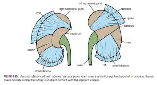
The kidneys are reddish brown and lie behind the peritoneum high
up on the posterior abdominal wall on either side of the vertebral column; they
are largely under cover of the costal margin.
The right kidney lies slightly lower than the left kidney because
of the large size of the right lobe of the liver. With contraction of the
diaphragm during respiration, both kidneys move downward in a vertical
direction by as much as 1 in. (2.5 cm). On the medial concave border of each
kidney is a vertical slit that is bounded by thick lips of renal substance and
is called the hilum. The hilum extends into a large cavity called the renal
sinus. The hilum transmits, from the front backward, the renal vein, two
branches of the renal artery, the ureter, and the third branch of the renal
artery (VAUA). Lymph vessels and sympathetic fibers also pass through the
hilum
Renal
Mobility
The kidneys are maintained in their normal position by
intraabdominal pressure and by their connections with the perirenal fat and
renal fascia. Each kidney moves slightly with respiration. The right kidney
lies at a slightly lower level than the left kidney, and the lower pole may be
palpated in the right lumbar region at the end of deep inspiration in a person
with poorly developed abdominal musculature. Should the amount of perirenal fat
be reduced, the mobility of the kidney may become excessive and produce
symptoms of renal colic caused by kinking of the ureter. Excessive mobility of
the kidney leaves the suprarenal gland undisturbed because the latter occupies
a separate compartment in the renal fascia.
Kidney
Trauma
The kidneys are well protected by the lower ribs, the lumbar
muscles, and the vertebral column. However, a severe blunt injury applied to
the abdomen may crush the kidney against the last rib and the vertebral column.
Depending on the severity of the blow, the injury varies from a mild bruising
to a complete laceration of the organ. Penetrating injuries are usually caused
by stab wounds or gunshot wounds and often involve other viscera. Because 25%
of the cardiac outflow passes through the kidneys, renal injury can result in
rapid blood loss
Kidney
Tumors
Malignant tumors of the kidney have a strong tendency to
spread along the renal vein. The left renal vein receives the left testicular vein
in the male, and this may rarely become blocked, producing left-sided
varicocele.
Renal
Pain
Renal pain varies from a dull ache to a severe pain in the
flank that may radiate downward into the lower abdomen. Renal pain can result
from stretching of the kidney capsule or spasm of the smooth muscle in the
renal pelvis. The afferent nerve fibers pass through the renal plexus around
the renal artery and ascend to the spinal cord through the lowest splanchnic nerve
in the thorax and the sympathetic trunk. They enter the spinal cord at the
level of T12. Pain is commonly referred along the distribution of the subcostal
nerve (T12) to the flank and the anterior abdominal wall.
Transplanted
Kidneys
The iliac fossa on the posterior abdominal wall is the usual
site chosen for transplantation of the kidney. The fossa is exposed through an
incision in the anterior abdominal wall just above the inguinal ligament. The
iliac fossa in front of the iliacus muscle is approached retroperitoneally. The
kidney is positioned and the vascular anastomosis constructed. The renal artery
is anastomosed end to end to the internal iliac artery and the renal vein is
anastomosed end to side to the external iliac vein. Anastomosis of the branches
of the internal iliac arteries on the two sides is sufficient so that the
pelvic viscera on the side of the renal arterial anastomosis are not at risk.
Ureterocystostomy is then performed by opening the bladder and providing a wide
entrance of the ureter through the bladder wall.


No comments:
Post a Comment