Inguinal
Canal
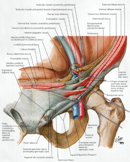
The inguinal canal is an oblique passage through the lower
part of the anterior abdominal wall. In the males, it allows structures to pass
to and from the testis to the abdomen. In females, it allows the round ligament
of the uterus to pass from the uterus to the labium majus. The canal is about
1.5 in. (4 cm) long in the adult and extends from the deep inguinal ring, a
hole in the fascia transversalis , downward and medially to the superficial
inguinal ring, a hole in the aponeurosis of the external oblique muscle . It
lies parallel to and immediately above the inguinal ligament. In the newborn
child, the deep ring lies almost directly posterior to the superficial ring so
that the canal is considerably shorter at this age. Later, as the result of
growth, the deep ring moves laterally. The deep inguinal ring,* an oval opening
in the fascia transversalis, lies about 0.5 in. (1.3 cm) above the inguinal
ligament midway between the anterior superior iliac spine and the symphysis
pubis . Related to it medially are the inferior epigastric vessels, which pass
upward from the external iliac vessels. The margins of the ring give attachment
to the internal spermatic fascia (or the internal covering of the round
ligament of the uterus). The superficial inguinal ring* is a triangular-shaped
defect in the aponeurosis of the external oblique muscle and lies immediately
above and medial to the pubic tubercle . The margins of the ring, sometimes
called the crura, give attachment to the external spermatic fascia.

Function of the
Inguinal Canal
The inguinal canal allows structures of the spermatic cord to pass to and from the testis to the abdomen in the male. (Normal spermatogenesis takes place only if the testis leaves the abdominal cavity to enter a cooler environment in the scrotum.) In the female, the smaller canal permits the passage of the round ligament of the uterus from the uterus to the labium majus
The inguinal canal allows structures of the spermatic cord to pass to and from the testis to the abdomen in the male. (Normal spermatogenesis takes place only if the testis leaves the abdominal cavity to enter a cooler environment in the scrotum.) In the female, the smaller canal permits the passage of the round ligament of the uterus from the uterus to the labium majus
Walls of the Inguinal Canal
Anterior wall:
External oblique aponeurosis, reinforced laterally by the origin of the internal oblique from the inguinal ligament . This wall is therefore strongest where it lies opposite the weakest part of the posterior wall, namely, the deep inguinal ring.
External oblique aponeurosis, reinforced laterally by the origin of the internal oblique from the inguinal ligament . This wall is therefore strongest where it lies opposite the weakest part of the posterior wall, namely, the deep inguinal ring.
Posterior wall:
Conjoint tendon medially, fascia transversalis laterally . This wall is therefore strongest where it lies opposite the weakest part of the
Conjoint tendon medially, fascia transversalis laterally . This wall is therefore strongest where it lies opposite the weakest part of the
anterior wall:
namely, the superficial inguinal ring. Roof or superior wall. Arching lowest fibers of the internal oblique and transversus abdominis muscles .
namely, the superficial inguinal ring. Roof or superior wall. Arching lowest fibers of the internal oblique and transversus abdominis muscles .
Floor or inferior wall:
Upturned lower edge of the inguinal ligament and, at its medial end, the lacunar ligament
Upturned lower edge of the inguinal ligament and, at its medial end, the lacunar ligament
Vas
Deferens (Ductus Deferens)
The vas deferens is a cordlike structure that can be
palpated between finger and thumb in the upper part of the scrotum. It is a
thick-walled muscular duct that transports spermatozoa from the epididymis to
the urethra.
No comments:
Post a Comment