Scrotum
The scrotum is an outpouching of the lower part of the anterior
abdominal wall and contains the testes, the epididymides, and the lower ends of
the spermatic cords
The wall of the scrotum has the following layers:
■■
Skin
■■
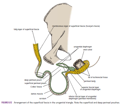
Superficial fascia; the dartos muscle, which is smooth muscle, replaces the
fatty layer of the anterior abdominal wall, and Scarpa’s fascia (membranous
layer), now called Colles’ fascia.
■■
External spermatic fascia derived from the external oblique
■■ Cremasteric
fascia derived from the internal oblique
■■
Internal spermatic fascia derived from the fascia transversalis
■■
Tunica vaginalis, which is a closed sac that covers the anterior, medial, and
lateral surfaces of each testis Because the structure of the scrotum, the
descent of the testes, and the formation of the inguinal canal are
interrelated
Blood
Supply
Subcutaneous plexuses and arteriovenous anastomoses promote
heat loss and thus assist in the environmental control of the temperature of
the testes.
Arteries
The external pudendal branches of the femoral and scrotal branches
of the internal pudendal arteries supply the scrotum.
Veins
The veins accompany the corresponding arteries.

Erection
of the Penis
Erection in the male is gradually built up as a consequence
of various sexual stimuli. Pleasurable sight, sound, smell, and other psychic
stimuli, fortified later by direct touch sensory stimuli from the general body
skin and genital skin, result in a bombardment of the central nervous system by
afferent stimuli. Efferent nervous impulses pass down the spinal cord to the
parasympathetic outflow in the second, third, and fourth sacral segments. The
parasympathetic preganglionic fibers enter the inferior hypogastric plexuses
and synapse on the postganglionic neurons. The postganglionic fibers join the
internal pudendal arteries and are distributed along their branches, which
enter the erectile tissue at the root of the penis. Vasodilatation of the
arteries now occurs, producing a great increase in blood flow through the blood
spaces of the erectile tissue. The corpora cavernosa and the corpus spongiosum
become engorged with blood and expand, compressing their draining veins against
the surrounding fascia. By this means, the outflow of blood from the erectile
tissue is retarded so that the internal pressure is further accentuated and
maintained. The penis thus increases in length and diameter and assumes the
erect position. Once the climax of sexual excitement is reached and ejaculation
takes place, or the excitement passes off or is inhibited, the arteries
supplying the erectile tissue undergo vasoconstriction. The penis then returns
to its flaccid state
Ejaculation
During the increasing sexual excitement that occurs during
sex play, the external urinary meatus of the glans penis becomes moist as a
result of the secretions of the bulbourethral glands
Friction
of the glans penis
reinforced by other afferent nervous impulses, results in a discharge along the sympathetic nerve fibers to the smooth muscle of the duct of the epididymis and the vas deferens on each side, the seminal vesicles, and the prostate. The smooth muscle contracts, and the spermatozoa, together with the secretions of the seminal vesicles and prostate, are discharged into the prostatic urethra. The fluid now joins the secretions of the bulbourethral glands and penile urethral glands and is then ejected from the penile urethra as a result of the rhythmic contractions of the bulbospongiosus muscles, which compress the urethra. Meanwhile, the sphincter of the bladder contracts and prevents a reflux of the spermatozoa into the bladder. The spermatozoa and the secretions of the several accessory glands constitute the seminal fluid, or semen. At the climax of male sexual excitement, a mass discharge of nervous impulses takes place in the central nervous system. Impulses pass down the spinal cord to the sympathetic outflow (T1 to L2). The nervous impulses that pass to the genital organs are thought to leave the cord at the first and second lumbar segments in the preganglionic sympathetic fibers. Many of these fibers synapse with postganglionic neurons in the first and second lumbar ganglia. Other fibers may synapse in ganglia in the lower lumbar or pelvic parts of the sympathetic trunks. The postganglionic fibers are then distributed to the vas deferens, the seminal vesicles, and the prostate via the inferior hypogastric plexuses.
reinforced by other afferent nervous impulses, results in a discharge along the sympathetic nerve fibers to the smooth muscle of the duct of the epididymis and the vas deferens on each side, the seminal vesicles, and the prostate. The smooth muscle contracts, and the spermatozoa, together with the secretions of the seminal vesicles and prostate, are discharged into the prostatic urethra. The fluid now joins the secretions of the bulbourethral glands and penile urethral glands and is then ejected from the penile urethra as a result of the rhythmic contractions of the bulbospongiosus muscles, which compress the urethra. Meanwhile, the sphincter of the bladder contracts and prevents a reflux of the spermatozoa into the bladder. The spermatozoa and the secretions of the several accessory glands constitute the seminal fluid, or semen. At the climax of male sexual excitement, a mass discharge of nervous impulses takes place in the central nervous system. Impulses pass down the spinal cord to the sympathetic outflow (T1 to L2). The nervous impulses that pass to the genital organs are thought to leave the cord at the first and second lumbar segments in the preganglionic sympathetic fibers. Many of these fibers synapse with postganglionic neurons in the first and second lumbar ganglia. Other fibers may synapse in ganglia in the lower lumbar or pelvic parts of the sympathetic trunks. The postganglionic fibers are then distributed to the vas deferens, the seminal vesicles, and the prostate via the inferior hypogastric plexuses.
Male
Urethra
The male urethra is about 8 in. (20 cm) long and extends from
the neck of the bladder to the external meatus on the glans penis. It is
divided into three parts: prostatic, membranous, and penile.
The prostatic urethra is described on page 278. It is about
1.25 in. (3 cm) long and passes through the prostate from the base to the apex.
It is the widest and most dilatable portion of the urethra. The membranous
urethra is about 0.5 in. (1.25 cm) long and lies within the urogenital
diaphragm, surrounded by the sphincter urethrae muscle. It is the least
dilatable portion of the urethra.
The penile urethra is about 6 in. (15.75 cm) long and is
enclosed in the bulb and the corpus spongiosum of the penis. The external
meatus is the narrowest part of the entire urethra. The part of the urethra
that lies within the glans penis is dilated to form the fossa terminalis
(navicular fossa). The bulbourethral glands open into the penile urethra below the
urogenital diaphragm.
No comments:
Post a Comment