Uterus
The uterus is a hollow, pear-shaped organ with thick
muscular walls. In the young nulliparous adult, it measures 3 in. (8 cm) long,
2 in. (5 cm) wide, and 1 in. (2.5 cm) thick. It is divided into the fundus,
body, and cervix.
The fundus is the part of the uterus that lies above the entrance of the uterine tubes.
The body is the part of the uterus that lies below the entrance of the uterine tubes.
The fundus is the part of the uterus that lies above the entrance of the uterine tubes.
The body is the part of the uterus that lies below the entrance of the uterine tubes.
The cervix is the narrow part of the uterus. It
pierces the anterior wall of the vagina and is divided into the supravaginal and
vaginal parts of the cervix. The cavity of the uterine body is triangular in
coronal section, but it is merely a cleft in the sagittal plane. The cavity of
the cervix, the cervical canal, communicates with the cavity of the body
through the internal os and with that of the vagina through the external os.
Before the birth of the first child, the external os is circular. In a parous
woman, the vaginal part of the cervix is larger, and the external os becomes a
transverse slit so that it possesses an anterior lip and a posterior lip.
Relations
■■
Anteriorly: The body of the uterus is related anteriorly to the uterovesical
pouch and the superior surface of the bladder. The supravaginal cervix is
related to the superior surface of the bladder. The vaginal cervix is related
to the anterior fornix of the vagina.
■■
Posteriorly: The body of the uterus is related posteriorly to the rectouterine
pouch (pouch of Douglas) with coils of ileum or sigmoid colon within it.
■■
Laterally: The body of the uterus is related laterally to the broad ligament
and the uterine artery and vein. The supravaginal cervix is related to the
ureter as it passes forward to enter the bladder. The vaginal cervix is related
to the lateral fornix of the vagina. The uterine tubes enter the superolateral
angles of the uterus, and the round ligaments of the ovary and of the uterus are
attached to the uterine wall just below this level.

Function
The uterus serves as a site for the reception, retention,
and nutrition of the fertilized ovum.
Positions of the Uterus
In most women, the long axis of the uterus is bent forward on
the long axis of the vagina. This position is referred to as anteversion of
the uterus. Furthermore, the long axis of the body of the uterus is bent
forward at the level of the internal os with the long axis of the cervix. This position
is termed anteflexion of the uterus. Thus, in the erect position and
with the bladder empty, the uterus lies in an almost horizontal plane. In some
women, the fundus and body of the uterus are bent backward on the vagina so
that they lie in the rectouterine pouch (pouch of Douglas). In this situation,
the uterus is said to be retroverted. If the body of the uterus is, in
addition, bent backward on the cervix, it is said to be retroflexed

Supports
of the Uterus
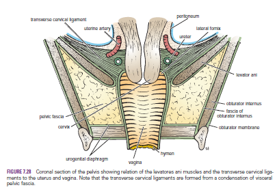
The uterus is supported mainly by the tone of the levatores ani
muscles and the condensations of pelvic fascia, which form three important
ligaments.
The
Levatores Ani Muscles and the Perineal Body
They form a broad
muscular sheet stretching across the pelvic cavity, and, together with the
pelvic fascia on their upper surface, they effectively support the pelvic
viscera and resist the intra-abdominal pressure transmitted downward through
the pelvis. The medial edges of the anterior parts of the levatores ani muscles
are attached to the cervix of the uterus by the pelvic fascia.
No comments:
Post a Comment